Soft Markers for Aneuploidy

Here’s this week’s RoshReview Question of the Week!

A 38-year-old G1P0 woman at 20 weeks gestation presents to the clinic for her anatomy ultrasound examination. She underwent a first-trimester screen, which showed a borderline nuchal translucency of 3.1 mm. Which one of the following isolated ultrasound findings confers the greatest risk for trisomy 21?

Check out the links above to see if you answered correctly. Also, you can enter for a chance to win a Rosh Review Qualifying Exam (“written boards”) QBank!


Check out the SMFM Consult Series 57 for excellent companion reading!

What are the ultrasound soft markers, and why do we care? 

  • In the era of cell-free DNA, you might ask: what is the utility of soft markers? Aren’t they poor predictors of aneuploidy?

    • Originally introduced to improve the detection of Down syndrome over that of just age-based or serum-based screening 

    • While it is true that each isolated soft marker may be poor predictors, if we see multiple soft markers, that does improve sensitivity  

    • There may also be some misunderstanding of soft markers seen on ultrasound, and so the purpose here is to review some of these soft markers in the setting of cfDNA and discuss next steps 

  • Remember: patient’s baseline risk should not limit screening options, and cfDNA should be offered to all per ACOG and SMFM 

What are the first steps when you see a soft marker?

  • Make sure that the soft marker is truly isolated - look for other soft markers, fetal growth restriction, or other anomalies 

    • If you feel that your office is not equipped to do this, can refer to MFM to have a level II ultrasound performed - this is of course a discussion with the patient, and not all patients will want further evaluation 

  • Look at the patient’s history: 

    • What is their baseline risk? (age, family history, history of aneuploidy) 

    • What are their previous aneuploidy screening results? Did they have any? 

  • Ok, so I see one of the soft markers, what do I do next?

    • First of all, have they had cfDNA?

      • Most of the time, there is not much to do after that (again: ISOLATED soft marker) 

      • This is because with cfDNA, the posttest probability of a common aneuploidy (ie. Trisomy 21) of negative cfDNA is very low - it is lowered by 300x for trisomy 21

        • Per the consult series, the residual risk of a 35-yo woman, whose age related risk of Down syndrome is 1/356 is reduced to <1/50,000 after a negative cfDNA result  

    • But what if they didn’t have cfDNA? 

      • If they have had negative serum screening, also ok, no need to do further testing at this time 

        • The detection rate of serum screening test for Down is still high, about 81%-99% depending on the test 

      • If no screening at all, counsel about noninvasive aneuploidy testing - not all patients will want screening 

    • Remember: there isn’t an established cut off residual risk when there is recommendation to do diagnostic testing 

      • Many labs will establish a cutoff of 1:250 or 1:300 

    • SMFM does not recommend diagnostic testing for aneuploidy only for evaluation of isolated soft marker following negative serum or cfDNA screening result 

The Soft Markers (all photo credit to Radiopedia)

Obstetrical Ultrasound

Sonographic nomenclature breaks down into three main types of ultrasound:

  1. Limited US: for a specific, singular purpose (i.e., viability, placental location, presentation, cervical length).

  2. Standard US: A more thorough examination, which requires a number of elements including:

    • Fetal presentation and number 

    • Amniotic fluid volume

      1. AFI (normal 5-25) or DVP (normal 2-8)

      2. SMFM recommends using DVP over AFI to diagnose oligo in the third trimester as using AFI leads to more interventions without improving perinatal outcomes 

    • Fetal Heart Rate / Rhythm

    • Placental location 

      1. Previa covers cervical os, either partially or wholly.

      2. Low lying placental edge is within 2cm of os.

        1. Partial or marginal previa is unfavored terminology.

      3. Suspicion for abnormal placentation for previa in setting of history of cesarean. With previa, history of 

        1. 1 prior cesarean = 3% risk of PAS

        2. 2 prior cesarean = 11% risk of PAS

        3. 3 prior cesarean = 40% risk of PAS

        4. 4 prior cesarean = 61% risk of PAS

    • Fetal biometry/anatomic survey.

    • Cervix and adnexa when clinically appropriate and when technically feasible. 

  3. Specialized US: A highly technical ultrasound that focuses on particular organ systems or uses dynamic measures.

    1. Examples include level 2 ultrasounds which provide further anatomic detail than a standard US, or fetal doppler ultrasonography, biophysical profiles, or fetal echocardiograms.

Important Facts for Ultrasound, by Trimester:

First Trimester: Know the absolute and relative criteria for early pregnancy failure, from SRU’s publication in NEJM:

(c) NEJM, 2013

2nd / 3rd Trimester: Know components of a basic growth scan by the Hadlock formula. Some representative photos are here! Images courtesy of www.fetalultrasound.com. Be sure to check out The OBG Project 2nd Trimester Ultrasound Atlas if you’re a chief resident with access to OBG First!

Appropriate plane for BPD and HC measurements

Appropriate plane for fetal abdominal circumference.

Appropriate plane for fetal abdominal circumference.

Appropriate plane and landmarks for fetal femoral length measurement.

Diagnostic Imaging During Pregnancy and Lactation

Today we’re going to review a source of constant consultation and confusion: diagnostic imaging during pregnancy and breastfeeding. ACOG CO 723 is the definitive reading on this subject, and we use it to structure this episode. Critical take home: ACOG states that critical imaging studies should not be withheld from a pregnant patient if needed to make a diagnosis. 

Ultrasound

  • Sonography utilizes sound waves to produce a visible image, and is not a form of ionizing radiation.

    • Thus, it is considered the safest mode of imaging in pregnancy.

    • However, ACOG still recommends sticking to the ALARA principle of exposure in pregnancy (“As Low As Reasonably Achievable”) to minimize any potential untoward effects. 

    • One of these theoretical effects involves color or spectral flow Doppler. Due to its intensity, the theoretical temperature increase surrounding the area being study can be as high as 2deg C, or 3.6deg F. It’s unlikely than any temperature increase would be sustained at any fetal anatomic site to cause harm. However, for this reason, even ultrasound exposure should be used judiciously. 

MRI

  • Allows for visualization of soft-tissue structures like ultrasound

    • However, as MRI is operator-independent, pick up rates for certain pathologies like appendicitis tend to be higher.

    • There are no special contraindications or considerations in pregnancy for non-contrast MRI, other than the usual screening surrounding metal or magnet-sensitive implants, such as pacemakers. 

  • Non-contrast MRI is sufficient for diagnosis; however, some diagnoses or studies may be improved by the use of gadolinium-based contrast, for which there is uncertainty regarding fetal effects.

    • Gadolinium is water-soluble, and thus crosses the placenta into fetal circulation.

    • Free gadolinium is toxic, so it is bound, or chelated, when administered for studies.

      • There is concern that since this bound gadolinium can enter fetal circulation, it can recycle in the fetal circulation. This potentially could sit for long enough that the gadolinium could dissociate and become free; thus become toxic. 

      • Given at least the concern for potential poor outcomes, gadolinium-based contrast should be limited in use to cases where there is an absolute clear benefit to its administration.

    • Gadolinium’s water-solubility makes it an OK contrast agent to use during lactation.

      • Less than 0.04% of a dose of gadolinium will be excreted in breastmilk in the first 24 hours, and less than 1% of this will be absorbed in the infant GI tract. Thus, breastfeeding should not be interrupted after gadolinium contrast studies.

CT, XRAY, and other ionizing radiation studies
Before talking about ionizing radiation studies, it’s important to know some vocabulary and measurements of radiation:

  • Exposure is the number of ions produced by radiation in the form of X-rays or gamma rays per kilogram of air. This is measured in Roentgen units.

  • Dose is the amount of energy deposited per kilogram of tissue. This is the usual consideration when we talk about radiation in pregnancy. This is measured in rads or in Gray units; 100 rad is equivalent to 1 Gray.

  • Relative effective dose is the amount of energy deposited per kilogram of tissue, and normalized for biological effectiveness on the tissue. This is measured in Roentgen equivalent men (rem) or Sievert units.


Again, the dose is what we usually consider and track with respect to radiation in pregnancy.

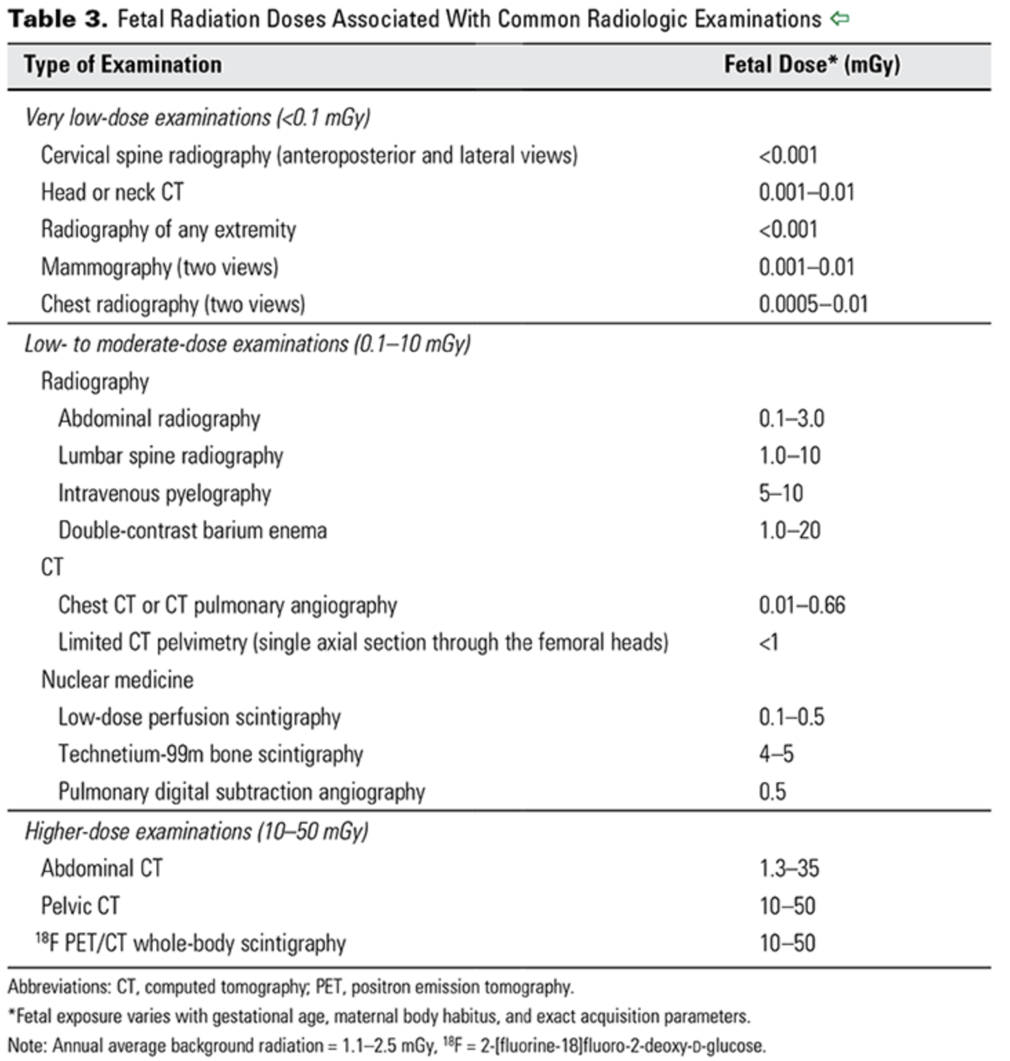
  • The background dose of radiation a fetus is exposed to during pregnancy is around 1 mGy.

  • From CO 723 — a reference for doses associated with different imaging studies.

ACOG CO 723

ACOG CO 723

The risk of radiation exposure on a developing fetus depends on both the dose of radiation, as well as the gestational age at which the exposure occurs.

  • For instance, if an exposure of 50-100 mGy occurs prior to implantation (0-2 weeks post fertilization), there is generally an all or none effect; that is to say, this usually results in miscarriage, or no consequence at all.

  • During organogenesis, or 2-8 weeks post-fertilization, congenital anomalies or growth restriction can be seen with cumulative doses of 200-250 mGy.

  • The risk of severe intellectual deficit or microcephaly is most prominent around 8-15 weeks, with doses between 60 - 300 mGy.

    • There is an estimated 25 point IQ loss per 1000 mGy exposure during this time period. 

    • A lower risk of severe intellectual disability may persist through 25 weeks gestation, though again with exposures of 250mGy or more.

  • Other risks include childhood cancer. With respect to leukemia, it is estimated the risk of childhood leukemia increases 1.5-2 fold with a 10-20mGy dose, over a background leukemia risk of 1 in 3000.

  • Radiologists and radiation physicists can help to calculate doses for patients exposed to multiple studies or with occupational hazards. 

With respect to contrast:

  • Oral contrast poses no real or theoretical harm to pregnant or lactating mothers and their infants.

  • IV contrast tends to be iodinated, but is also water-soluble.

    • So similarly to gadolinium, in pregnant patients this crosses the placenta.

    • Animal studies have demonstrated no teratogenic effects from its use, but it is recommended to limit use of iodinated contrast unless necessary.

    • Also similarly to gadolinium, because of this water solubility, iodinated contrast is excreted minimally in breastmilk, and breastfeeding should be continued without interruption.

Nuclear medicine studies

Radioisotopes for nuclear medicine studies, such as VQ scans, thyroid scans, and bone scans, are variable in their potential effects on the fetus.

  • Technetium-99 is one of the most common radioisotopes used for these studies, and given its short half life of 6 hours as well as its pure gamma ray emission, is generally accepted as safe to use when indicated in pregnancy.

  • Radioactive iodine (I-131), by contrast (punny!), readily crosses the placenta and has a half-life of 8 days, and has known adverse effects on the fetal thyroid. Thus, it is contraindicated for use in pregnancy, and is also recommended against use in breastfeeding mothers until breast milk has been cleared of the radioisotope.