Espresso: Umbilical Cord Gas Interpretation

Building somewhat on our fetal circulation episode from last week, today we’ll talk about umbilical cord gases. From an obstetrics perspective, these can be challenging to really interpret, but the simple interpretation is often worth some CREOG points if you can analyze these systematically.

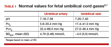
Remember, the umbilical vein is carrying oxygenated blood, and the umbilical arteries are carrying deoxygenated blood. This can help you remember the normal values, as they’ll be opposite those for an ABG versus VBG on an adult. Additionally, the umbilical vein is originating at the site of the placental interface with the mother -- so venous pHs will give a sense of maternal acid-base status, or the acid-base status at this interface. For this reason, the arterial pH is more helpful to truly measure fetal acid-base status.

The components of the blood gas are:

  • pH: represents the inverse log of the concentration of hydrogen ions in the circulating blood, or how acidic the blood is. In essence, more acid represents a lower pH, which represents more compromise. 

    • Normal value for a venous pH is around 7.35 (as it is in adult blood).

    • Normal value for an arterial pH is around 7.28.

  • pO2: the pressure of oxygen (in essence its concentration) in fetal blood.

  • pCO2: similarly, the pressure/concentration of CO2 in fetal blood.

    • The pO2 and pCO2 can given additional clues to help with non-straightforward (i.e., mixed) acidosis.

  • Base Excess/Deficit: in blood, acid is buffered by bicarbonate ions. The base excess or deficit represents how much difference there is between those bicarbonate ions and hydrogen ions in order to return to a normal pH value of 7.35 in the umbilical vein. An excess is more bicarb; a deficit is less bicarb. However, these tend to get used interchangeably, and in these acid-base status questions, you’ll see the “excess” written as a negative number — implying what is actually a deficit.

    • Normal values for base excess are around 4 mmol/L in both the umbilical artery and vein.

    • A more significant base deficit signifies a metabolic acidosis -- i.e., the process causing insult has been longstanding, and there has been time to utilize bicarbonate to buffer the acid. 

    • A lower base deficit signifies a respiratory acidosis -- i.e., the process has been acute, so there has been no buffering of the hydrogen ions. 

      • A base deficit of 12 mmol/L has been suggested as severe, and more suggestive of metabolic acidosis. 

(c) MDEdge

(c) MDEdge

What about administering more O2 to the mother? Won’t that help things and reduce the fetal risk of acidosis?

If only it were that simple! Sadly, the answer is no. In most cases, maternal hemoglobin is fully saturated on room air. Fetal hemoglobin has a greater O2 avidity, and will pull O2 across the placental circulation. But when maternal blood is already saturated, the fetus won’t get any more O2 even if you pump it up to 4000L a minute by mega face mask! Some studies have suggested the additional free O2 in maternal serum may actually lead to vasospasm and cause harm! 

The exception to this certainly is a change in maternal oxygenation or an indication for maternal O2 use -- but these indications suggest that maternal Hb is less than 100% saturated. 

When should I get a cord gas?

It’s a good idea to practice the technique for cord gas collection, which requires collecting a 10-20cm doubly-clamped (i.e., proximally and distally) cord segment. Even on routine, vigorous deliveries, getting into this habit as part of your deliveries will help you be prepared. 

Cord gases are not recommended to be sent with delayed cord clamping, so don’t send these if DCC is part of your practice! However, collecting the cord segment can be good practice for those learning proper technique.

There are no consensus rules about when to send a cord gas sample. At our institution, the general thought is “if you think you need one, send one.” However, common scenarios where cord gas sampling can be helpful to at least set aside on a “just in case basis”  include:

  • Nonvigorous infant at delivery (i.e., Apgars at 5 mins less than 7)

  • Category III or “bad category II” tracings

  • Operative deliveries performed for NRFHT

  • Multiple gestation

  • Premature infants

  • Meconium stained fluid

  • Growth restriction

  • Breech deliveries

  • Shoulder dystocia

  • Intrapartum fevers or chorioamnionitis

Obviously this list is non-exhaustive, but goes to show there are a lot of indications! Some literature has suggested even universal arterial blood sampling at delivery may be cost-effective. 

The best way to learn this is to do some practice cases. Check out the below resources for some practice questions and further explanations.




Fetal Circulation

One of the neonatology/pediatric points the CREOG exam will test on is flow of blood through the fetal circulation. It can be quite confusing, but it’s worth remembering. We’ll take you on the journey of a red blood cell in today’s episode.

The important foundational bit of knowledge for this is the nomenclature of arteries and veins. Arteries carry blood away from the heart (Arteries Away), whereas veins carry blood towards the heart. Arteries and veins do not denote oxygenation status, particularly in the fetal circulation!

(C) Children’s Hospital of Philadelphia

Let’s start at the umbilical vein, which is carrying oxygenated blood from the placenta towards the fetal heart. Remember there is a single large umbilical vein with normal umbilical cords.

  • The umbilical vein enters at the umbilicus, and moves superiorly towards the liver, where it ultimately needs to meet the inferior vena cava. However, the umbilical vein naturally empties into the portal hepatic vein

    • This is where we encounter our first fetal shunt, the ductus venosus.

      • This allows oxygenated blood from the umbilical vein to connect to the inferior vena cava, bypassing the portal vein and the liver. 

      • The ductus venosus closes functionally in term infants within minutes of birth, and full closure naturally occurs within one week of birth. In preterm infants this may take longer. The remnant structure is known as the ligamentum venosum.

  • From the IVC, we can get blood into the right atrium of the heart. Now blood will move to the right ventricle naturally in adult circulation. In fetal circulation, though, the lungs have yet to open. The pulmonary circulation is of very high resistance. Rather than take the long, high resistance trip around the lungs, we encounter our second shunt, the foramen ovale between the right and left atria. 

    • The relatively high pressure in the right atrium allows for blood to move across this shunt into the left atrium. 

    • With the first neonatal breaths, the lungs open and the resistance to the pulmonary circulation drastically drops. This allows for the foramen ovale to close, as the septum secundum (some tissue in the right atrium where the foramen ovale is located), is effectively a one way valve from right to left; when flow starts to go left-to-right, this valve closes.

      • In up to 25% of adults, this one-way valve closure is not completely effective, leading to the patent foramen ovale.

  • Now blood is in the left heart, where it can move from left atria, to left ventricle, to aorta, and now supply the fetal brain and other tissues.

  • However, some blood may still move to the right ventricle in spite of the pressure gradient, and try to move through the pulmonary circulation. 

    • To exit the pulmonary circulation more quickly and supply oxygenated blood to the lower extremities, we encounter our third shunt, the ductus arteriosus. This connects the pulmonary artery to the descending aorta. After birth, this closes and becomes the ligamentum arteriosum

    • In some individuals, the ductus arteriosus remains open, leading to a patent ductus arteriosus. Because of the change in pressure after birth, now oxygenated blood is leaving the aorta and overloading the pulmonary artery. This can lead to pulmonary hypertension and right heart failure. 

      • In PDA and rarely PFO, but more commonly with ventricular septums, the pulmonary hypertension becomes so great as to change the pressure differential again (i.e., pulmonary or right heart circulation pressure is greater than left heart or systemic circulation). This changes the shunt to send deoxygenated blood from the right heart into the systemic circulation, and is known as Eisenmeger syndrome

  • Now that we’ve gotten all the blood to the left heart, it moves through the arteries to supply organs and tissues, and will end up in the veins. 

    • Coming from superiorly, blood will end up in the superior vena cava, and end up back in the right atrium. From here, it’s the same cycle all over again -- some will go through the foramen ovale, some will go to the right ventricle and pass through the ductus arteriosus. 

    • If blood went inferiorly (i.e., went through the descending aorta/ductus arteriosus), the umbilical arteries will carry blood back towards the placenta for re-oxygenation and deposition of CO2 and waste products. 

      • The umbilical arteries originate off the internal iliac arteries bilaterally. After birth, they become obliterated and are known as the medial umbilical ligaments. These can be seen during laparoscopic surgery and are good markers for the position of the superior vesical arteries. More on that in a future episode on pelvic anatomy!

Though we have the anatomic picture above, some folks may find a schematic helpful. Run through this a few times before your exam and you’ll be golden!

Trial of Labor after Cesarean (TOLAC)

In 2016, the US cesarean delivery rate was 31.9%. With ever increasing volumes of cesarean delivery, TOLAC has become a popular option for patients desiring vaginal delivery. On today’s episode, we examine TOLAC and share some counseling pointers in thinking about your patient. ACOG PB 205 is the requisite reading for the topic.

While there are no RCTs comparing TOLAC to planned cesarean, the relative benefits are easy to see: there is less recovery time, the patient avoids major surgery, and the potential sequelae of complications from major surgery — worsened bleeding, more opportunity for infection, more risk of complications requiring additional procedures. However, TOLAC is not without risk. We primarily counsel with respect to uterine rupture. Evaluations of “rupture” though have varied in the literature; it’s important to keep a discerning eye, as what is classified as rupture in some series is very different than what is in others. ACOG suggests the rate of uterine rupture in a patient with one low transverse cesarean delivery is around 0.5 - 0.9 %. Otherwise, maternal risks are fairly equal. Neonatal risks are also considered fairly equal, though with some increased risk associated with TOLAC.

ACOG PB 205

ACOG PB 205

We can think about patients who should be counseled against TOLAC:

  • Those at high risk of uterine rupture: ie. those with classic uterine incision, T-incision, prior uterine rupture, or extensive prior uterine fundal surgery like a myomectomy.

  • Women who are not otherwise candidates to have vaginal deliveries: ie. previa.

  • Women who desire homebirth: While ACOG does not definitely say that you cannot TOLAC in this instance, if you don’t access to emergency cesarean delivery, it is recommended that these patients have a discussion regarding the hospitals resources and possibly referral to a hospital that does have access to emergency cesarean delivery.

We can also consider patients for whom there may be a question of whether TOLAC is appropriate:

  • Low vertical incision? 

    1. Few studies, but those that have looked at them have shown similar rates of vaginal deliveries as low transverse. Can consider TOLAC!

  • Twins? 

    1. Studies show similar rates of successful VBAC in twins as in singleton gestations 

  • Obesity 

    1. Unfortunately, higher BMI seems to have an inverse relationship with success of VBAC. 85% of normal weight women achieve VBAC while only 65% of morbidly obese women do. However, morbidly obese women also can have more complications with an elective repeat cesarean, so counseling should be individualized

  • Induction and augmentation of labor 

    1. Mechanical dilation can be used - ie. cervical foley 

    2. Misoprostol has been shown to have increased risk of uterine rupture, so should not be used in term patients who have had c/s or other major uterine surgery for induction 

    3. However, in women undergoing second trimester labor inductions (ie. for missed abortion, induction of labor for stillbirths), use of prostaglandins have shown similar results in women who have had scars on their uterus and those without; so these women can still have prostaglandins, especially because no fetal considerations 

  • What if they’ve had a uterine rupture? 

    • If the site of rupture or dehiscence is in the lower part of the uterus, their risk of uterine rupture in labor is 6%. If it is in the upper segment of the uterus, the rate of dehiscence in labor is up to 32%. While there is no high quality data to guide this, recommendations are generally for subsequent pregnancies to be delivered by cesarean between 36-37 weeks.

Counseling should be individualized, and the MFMU has excellent calculators to help guide you and your patients to a decision about TOLAC:

(not in labor) https://mfmunetwork.bsc.gwu.edu/PublicBSC/MFMU/VGBirthCalc/vagbirth.html

(at admission) https://mfmunetwork.bsc.gwu.edu/PublicBSC/MFMU/VGBirthCalc/vagbrth2.html

Considerations for Planned Singleton Breech Vaginal Delivery

Today’s episode dives more into the realm of expert opinion rather than hard science, and we hope some of our listeners will share their own experiences and criteria for offering planned breech labor!

ACOG CO 745, in addition to PB 161 on external cephalic version, deal with this topic, but there is much variation in the literature available. In particular, international guidelines on this topic are rather variable. The ObG Project has a great summary and links to these varying resources that is worth checking out.

There are particular risks to breech labor, and experienced provider hands are necessary, which is why almost 90% of planned term breech birth in the USA is performed by cesarean section. The 2000 Term Breech Trial, a multicenter randomized trial, noted perinatal morbidity and mortality was overall reduced with planned cesarean delivery than with planned vaginal delivery of term breech (1.6% vs 5.0%), with no differences in reported maternal morbidity or mortality. Follow up studies to the Term Breech Trial, however, have noted no differences in maternal or neonatal outcomes at 2 years.

Additional studies performed since this time have been mixed. While some prospective studies demonstrated excellent maternal and neonatal outcomes, both short- and long-term, they utilized very strict criteria and protocols for the selection of candidates offered a trial of breech labor. Cohort studies of breech birth in general populations demonstrates at least short-term risk of neonatal morbidity, including birth injury, nerve injury, and need for assisted ventilation. This risk is present with any trial of breech labor, including if intrapartum cesarean is performed, versus planned cesarean delivery.

Below is a sample protocol based on some of these studies with stricter inclusion criteria. We recognize there is likely some significant debate to be had on these criteria, and in particular clinical scenarios, so be sure to discuss with experienced obstetricians in your area as well as check your hospital’s own breech birth protocol.

(c) CREOGs over Coffee, 2019. Adapted from Hofmeyr/UpToDate, 2019.

Finally, intrapartum management should proceed according to usual obstetric practice. However with breech presentations, providers should closely consider a number of factors outlined below. Notably, these factors are largely based on expert opinion and guidelines from international societies.

  • Avoidance of early amniotomy, and preference for spontaneous rupture of membranes.

  • The progress of labor in the active phase, and progress of descent during active pushing. 

    • Cesarean delivery should be recommended with a protracted labor course, particularly in the active phase, as this may be indicative of fetopelvic disproportion. 

    • Use of oxytocin in the active phase of labor is discouraged.

    • With the achievement of full cervical dilation, the breech should reach the pelvic floor.

    • Passive descent should not be permitted for more than 90 minutes after achieving full cervical dilation.

    • With onset of active pushing, delivery by cesarean should be considered if the infant has not delivered within 30-60 minutes.

Cardiac Arrest in Pregnancy

Today we discuss a topic that we hope you never encounter, but want every OB, EM, and really any other person or medical professional to be prepared for cardiac arrest in pregnancy. The American Heart Association (AHA) Scientific Statement on Cardiac Arrest in Pregnancy can be found here and is essential companion reading.

(c) AHA

In preparation for a maternal cardiac event, a cesarean delivery kit should be available as part of the adult code cart. This at minimum should have a scalpel (#10 blade), betadine splash prep, clamps for cutting the umbilical cord, sponges, absorbable suture, and additional clamps and/or retractors if feasible. A neonatal resuscitation cart should accompany the adult cart if a maternal code is ongoing.

BLS is not different from standard for any other adult resuscitation, except for one key component: leftward displacement of the uterus. This allows for improved venous return to the right heart via the inferior vena cava, which may be compressed to some degree as early as 12 weeks gestation. Otherwise hand positioning, compression technique, and ventilation considerations in the BLS portion do not have any differences.

The ACLS algorithm also proceeds as usual, with the notable exception being performance of resuscitative hysterotomy (aka, peri-mortem cesarean section) at 4 minutes of pulseless arrest. This should be performed at any gestation above 20 weeks (i.e., fundal height at or above the umbilicus). It serves the dual purpose of improving maternal venous return, as well as protecting the fetus from consequences of prolonged anoxia.

Otherwise, ACLS algorithms use the same medications and doses, the same indications for shocks, and actually many times the same etiologies for arrest. However there are some pregnancy-specific considerations all physicians should recall, in a simple mnemonic:

(c) Society of Obstetric Anesthesia and Perinatology