Perinatal Mental Health, feat. Dr. Tiffany Moore-Simas and Dr. Nancy Byatt

Today on the podcast, we’re addressing perinatal mental health. While we’ve talked about depression on the show before, there’s so much more in this sphere as we’ll discuss today.


Joining us are two experts in this field who share their passion for this work with us. Dr. Tiffany Moore Simas is Chair and Professor of OB/GYN at UMass Memorial Health and UMass Chan Medical School as well as co-Chair of the ACOG Maternal Mental Health Expert Work Group. And Dr. Nancy Byatt is a tenured Professor of Psychiatry and OB/GYN at UMass Memorial Health and UMass Chan Medical School. Both serve as senior leaders with the Massachusetts Perinatal Psychiatry Access Program, MCPAP for Moms, and Lifeline For Moms.


Importance of Perinatal Mental Health

  • Mental health conditions are the most common complications of pregnancy – 1 in 5!

    • More common in adolescents, veterans, marginalized populations (BIPOC, poverty).

  • Untreated mental health conditions carry both short- and long-term consequences that can affect whole family:

    • o   Less engagement in medical care

    • o   Smoking, substance use

    • o   Preterm delivery, low birth weight, NICU admission

    • o   Lactation challenges, bonding issues

      • Parent with untreated mental health disorder is considered an Adverse Childhood Experience (ACE) for the infant.

    • o   Adverse partner relationships

  • Mortality: leading cause of preventable maternal mortality.

    • 100% of maternal deaths due to mental health, including suicide, overdose, are preventable!

  • Underdetected and undertreated

  • OB/GYNs can screen and help manage mental health conditions. The majority (80%) of depression, for example, is managed by primary care providers, not psychiatrists. As obstetric care clinicians, we are the primary care providers to pregnant and postpartum individuals and thus, we should be providing mental health care!

Screening for Perinatal Mood and Anxiety Disorders

  • In this context, perinatal refers to during pregnancy and the first year after pregnancy ends

  • Perinatal Mood and Anxiety Disorders primarily include depression, bipolar disorder, and anxiety or anxiety-related conditions (generalized anxiety disorder, PTSD, OCD).

  • Screens should be performed with validated tools that query the last 7-14 days of symptoms for anxiety and depression.

    • o   Validated tools:

      • PHQ-9, EPDS (depression)

      • GAD-7 (generalized anxiety)

    • o   ACOG recommends screening patients at least once during the perinatal period for depression and anxiety symptoms. If a patient is screened during pregnancy, additional screening should occur during the comprehensive postpartum visit.

      • We recommend screening: new OB visit, later in pregnancy (i.e., 3rd trimester) and postpartum given the almost even distribution of onset predating pregnancy, onset in pregnancy, and onset postpartum.

    • o   Data suggests that early detection and treatment improves outcomes.

  • Bipolar disorder screening:

    • o   In one study, 1 in 5 patients screening positive for postpartum depression actually had bipolar disorder.

      • Recall: bipolar disorder can worsen with antidepressant treatment (unopposed SSRIs) – thus, need to screen for bipolar before initiating pharmacotherapy and ideally universally to prevent harm!

    • o   Patients with bipolar disorder have higher risk of postpartum psychosis

      • Rare: 1-2/1000 perinatal individuals; but 70% have bipolar disorder!

      • 4% risk of infanticide with postpartum psychosis

      • This is a psychiatric emergency.

        • Often occurs within the first days of delivery and most cases occur within the first 3 weeks.

    • o   Screening options:

      • Mood Disorder Question (MDQ) – self administered

      • CIDI – clinician administered with branching logic

    • o   Appropriate to refer to psychiatry if bipolar disorder is suspected – more on resources to help later!

Positive Screening   General Principles of Treatment

  • Just like a glucola, our questionnaires for mental health concerns are screening tests. Subsequent assessment is critical to confirm diagnosis.

    • o   See resources collection at the end of these notes for help!

  • For depression and anxiety, there are three pillars of treatment:

    • o   Psychotherapy

    • o   Pharmacotherapy or medication

      • o   Adjunctive interventions

  • Treat based on level of severity. For information on assessing and treating perinatal mental health conditions, visit the ACOG website.

  • If pharmacotherapy is indicated/started, patients may have some concerns:

    • o   Provide reassurance

    • o   Frame risk/benefit discussion in treated disease vs. untreated disease as not treating is associated with risks - just like any other disease!

    • o   Use lowest effective dose and monotherapy when able

  • Find more information on educating patients about treatment on ACOG’s website.

Concerns for Suicidality or Harm To Baby

  • These can represent urgent clinical scenarios and further assessment and response is critical:

    • o   Thoughts of harming self or baby are common yet not all are necessarily a psychiatric emergency.

    • o   When assessing for risk of harm to self or others it is important to assess:

      • Ideation – Do they have thoughts of harming themselves or someone else?  Are the thoughts fleeting or do they persist?

      • Intent – Are they intending to act on it? Have they thought of how they could do harm themselves or someone else or die by suicide?

      • Plan - Are they planning to act on it?  Have they developed a plan for how to die by suicide or to harm someone else?

    • o   If you are concerned that the patient is at risk of harm to self or others, then it is important to obtain further assessment which includes an evaluation for whether the patient may need psychiatric hospitalization

    • o   Regardless of whether these are a psychiatric emergency, the presence of thoughts of harming self or baby are indicative of higher illness severity.

  • More information on ACOG’s website.

 Resources for Integrating Perinatal Mental Health Care into Your Practice


Sickle Cell Disease, feat Dr. David Abel

Here’s the RoshReview Question of the Week:

Which of the following is a precipitating factor for a painful crisis in a pregnant woman with sickle cell disease?

Check out the answer at the links above and get a sweet deal on RoshReview!


Today we’re joined once again by Dr. David Abel, an assistant professor in the Department of Obstetrics and Gynecology at Oregon Health Sciences University. Dr. Abel has previously joined us to talk about thalassemias and von Willebrand’s disease — he shares his passion for blood disorders again with us today to talk more on sickle cell!

Listen to our last sickle podcast here. 

Epidemiology of Sickle Cell Disease (HbSS)

  • Most common inherited hemoglobinopathy in the United States, and in fact is the most common inherited disease worldwide.

    • Autosomal recessive fashion — both parents need to be carriers.

  • Affects approximately 10 million people worldwide and approximately 100,000 people in the United States.

    • This translates into a prevalence of about 1 in 375 who have the disease.

  • Predominantly affects people of African ancestry

    • Two thirds of those that are affected reside in West Africa.

    • 50% of children born with sickle cell disease are born in Nigeria.

  • Sickle Cell Trait: 1 in 12 are carriers.

    • In the United States, approximately 7% to 9% of the African American population.

    • As the gene is widely distributed, other populations may be affected including those residing in the areas of the Mediterranean, Caribbean, South and Central America, as well as East India.

  • More than 90% of children with sickle cell disease in the United States survive into adulthood.

    • Compared to the general population, however, their lifespans are two or threedecades shorter and limited by both acute and chronic morbidity.

  • Sickle cell trait confers a survival advantage in malaria-endemic regions such as in sub-Saharan Africa where almost 80% of individuals with sickle cell anemia live — resulting in “positive screening” with respect to human evolution.

Hemoglobin Structure in Sickle Cell and Pathophysiology

  • Remember: thalassemias represent quantitative defects globin synthesis. By contrast, hemoglobin S is characterized by a qualitative defect of the beta globin gene.

    • HbS results from a single nucleotide substitution, an adenine- to-thymine substitution in the sixth codon of the beta globin polypeptide which replaces glutamic acid with valine.

      • From this one amnio acid change, rather than forming tetramers, under conditions of low oxygen tension, this hemoglobin S forms long inflexible chains or fibers.

      • They distort the red blood cell membrane, resulting in this sickled shape.

      • These distorted red blood cells are destroyed by the reticuloendothelial system, resulting in a moderate to severe anemia.

        • Compared to the normal life span of a red blood cell of 120 days, the life span of these sickled red blood cells is reduced to an average of 15 days.

  • These distorted red blood cells clog up the microvasculature —> obstruction and local ischemia which clinically manifests as a vasoocclusive crisis.

    • Repeated vasoocclusive crises can lead to interruption of normal perfusion of multiple organs, including the spleen, lungs, kidneys, heartand brain.

    • Adults with sickle cell disease are essentially functionally asplenic — increased incidence and severity of infection in patients with sickle cell disease.

  • Sickled red cells are also prone to lysis which releases free hemoglobin.

    • Damages the endothelium and may also lead to thrombosis.

    • Also consumes nitric oxide, an important vasodilator and thus can lead to an exacerbation of the ischemia.  

Testing for Sickle Cell Disease and Trait

  • Foundation of screening: CBC, Hb electrophoresis. High suspicion in patients with family history / ancestry and MCV < 80.

    • HbSS (disease) electrophoresis:

      • 85-95% hemoglobin S

      • Remaining mostly hemoglobin F, small component of hemoglobin A2

    • HbSB (trait) electrophoresis (assuming normal second beta gene):

      • 50-60% hemoglobin A (normal adult Hb)

      • 35-45% hemoglobin S

      • Small amounts hemoglobin F, hemoglobin A2

  • There are many other sickle genotypes:

    • Homozygous hemoglobin SS constitutes about 70% of these genotypes.

    • Hemoglobin C, which differs from hemoglobin S only in that the amnio acid lysine instead of a valine replaces glutamic acid in the beta globin gene, can exist in combination with hemoglobin S, thus is called hemoglobin SC disease.

    • Hemoglobin S may coexist with beta-thalassemia.

      • Hemoglobin S beta thalassemia zero is also identified as sickle cell anemia and is just as severe as hemoglobin SS.

      • Hemoglobin S beta thalassemia plus is not as severe as there is some hemoglobin A that is preserved.

Maternal and Fetal Considerations with Sickle Cell

  • During pregnancy, the increase in red blood cell mass that normally occurs does not in those with sickle cell anemia.

    • 50%–70% of pregnancies with sickle cell disease require at least one hospitalization

    • 30%–40% will require a transfusion.

    • In the United States, the maternal mortality rate is approximately 10 times higher than it is for patients without sickle cell disease.

  • Vasoocclusive Crisis / Pain Crisis is most common cause of recurrent morbidity.

    • Can be precipitated by such factors as cold, physical exertion, dehydration and stress.

    • Opioids are a mainstay of treatment for a pain crisis — it is important not to withhold treatment for these patients.

  • Acute chest syndrome severe life-threatening form of a vasoocclusive crisis

    • Presents similarly to pneumonia.

      • Fever, tachypnea, chest pain, hypoxia and infiltrates noted on chest x-ray.

      • In addition to infectious agents, acute chest syndrome may also result from fat emboli, intrapulmonary aggregates of sickled red blood cells, atelectasis or pulmonary edema.

    • Patients with a history of frequent hospitalizations and/or episodes of acute chest syndrome correlate with increased risks during pregnancy.

    • The treatment of acute chest syndrome typically consists of antibiotics, usually ceftriaxone and azithromycin, pain control, and if needed oxygen and transfusion.

  • Other complications

    • Stroke: occurs in almost 25% by the age of 25

    • Splenic sequestration / asplenia

    • Acute renal failure

    • Acute cholecystitis

    • Pulmonary hypertension: 6 to 11% of patients with sickle cell disease

    • Venous thromboembolism: ocurs in 10-25% of those with sickle cell disease by age 40

      • In pregnancy risk elevates: 2x increased risk of stroke, 5x increased risk of cerebral vein thrombosis, 2x increased risk of pulmonary embolism, 2.5x increased risk of deep vein thrombosis.

    • Maternal infection complications: asymptomatic bacteriuria, pyelonephritis, sepsis and an almost ten-fold increased risk of pneumonia.

  • Placental Consequences:

    • Placental hypoperfusion with endothelial damage is the main contributor to adverse pregnancy outcomes.

    • Increased risk of preeclampsia and eclampsia, placental abruption, antepartum bleeding and alloimmunization.

  • Fetal consequences:

  • 2x increased risk of preterm birth

  • 3x risk of small-for-gestational age

  • 4x increased risk of stillbirth.

    • Serial fetal growth assessments and antepartum testing are needed.

  • Possible increased risk of neonatal abstinence syndrome due to the use of opioids to treat pain crises.

Should transfusion be used prophylactically?

  • 2015 meta-analysis of 12 observational studies with almost 1300 patients demonstrated a reduction in both maternal and perinatal mortality as well as a reduction in pain events and preterm birth.

  • 2016 Cochrane review that included only randomized controlled trials did not demonstrate a benefit with prophylactic when compared with selective transfusion.

  • Management strategy:

    • CBCs should be checked frequently, and a goal of maintaining a hemoglobin around 10 (same might say up to 12) and a percentage of hemoglobin S less than 35 to 40% is reasonable.

    • It is important to avoid iron overload when considering transfusion therapy which may lower target Hb for some individuals.

Treating Sickle Pain Beyond Opioids

  • Amitriptyline

  • Gabapentin

  • Selective serotonin reuptake inhibitors (SSRIs) and selective norepinephrine reuptake inhibitors (SNRIs)

  • Complementary (CAM) therapies

  • Hydroxyurea

    • Mainstay of treatment in the non-pregnant patient, as it reduces the risk of a vasoocclusive crisis and acute chest syndrome, thereby leading to improved survival and quality of life.

      • Patients with sickle cell disease who have higher amount of fetal hemoglobin tend to do better, and hydroxyurea increases fetal hemoglobin as well as reduces red blood cell adhesion and increases nitric oxide, the vasodilator we discussed earlier.

    • Has been found to cause birth defects in animals, although it has not been found to increase the risk of birth defects in humans. Still, it is generally avoided during pregnancy.

 Preconception Counseling for Sickle Cell

  • Discuss the increased risk of both maternal and fetal complications, though many can have a successful pregnancy.

    • Know history — past need for transfusions, the frequency of hospitalizations due to vasoocclusive crises and if there is a history of acute chest syndrome.

  • Due to the possibility of iron overload due to multiple transfusions, it is important to check a ferritin prior to prescribing iron.

  • ACOG recommends 4 mg of folic acid daily.

  • Screen for hypertension and treat if warranted to maintain a blood pressure less than 140/90.

  • A dilated eye examination performed by an ophthalmologist should be performed if not done within the past year.

  • If there are any concerns for nephropathy, refer to nephrology — screen for proteinuria.

  • If there are any concerns for pulmonary hypertension, an echocardiogram is recommended with cardiology referral if needed.

  • As most patients are functionally asplenic, pneumococcus, haemophilus influenza type b, and meningococcus immunizations are recommended.

  • Check antibody screen — possible risk of alloimmunization.

Cure for Sickle Cell Disease?

  • Have been accomplished with hematopoietic stem cell transplantation.

    • In this case, the donor may be related or unrelated.

  • Stem cell transplantation offers a cure, but can result in death, graft rejection, graft versus host disease, and sterility due to chemotherapy.

  • Gene therapy for sickle cell disease, where patients receive their own genetically modified hematopoietic stem cells, is still experimental and there are several clinical trials underway.  

Final Important Points on Managing Sickle Disease in Pregnancy

  • Hydroxyurea should be discontinued if it has not been already.

    • There are some who might consider restarting this after the fetal anatomical survey shows no evidence of abnormalities.

  • Chelation agents should also be discontinued.

  • Low-dose aspirin for preeclampsia risk reduction is recommended.

  • Monthly urinary cultures monitoring for asymptomatic bacteriuria and watching for any signs or symptoms of pyelonephritis.

    • Also reasonable to do in patients with sickle trait as risk is also increased in that population.

  • Frequent CBC monitoring, usually monthly is reasonable.

  • Interval fetal growth assessments every 3-4 weeks are recommended.

  • Antepartum testing starting at 32 weeks is indicated, with a delivery goal of 37 to 39 weeks.

  • Watch for preeclampsia is very important.

  • If a cesarean delivery is required, a preoperative transfusion may be prudent to increase hemoglobin levels to 8 to 10 g/dl.

  • Thromboprophyalxis: SCDs definitely; anticoagulation should be individualized.

    • Assuming the patient did not have a thromboembolic event during the pregnancy, could consider prophylactic low-molecular weight heparin for six weeks postpartum.

Dermatoses of Pregnancy, feat. Dr. Laura Hanks

Here’s the RoshReview Question of the Week!

A 30-year-old G1 woman presents at 34 weeks gestation to the office with severe itching and a rash that suddenly developed across her abdomen starting around her umbilicus. The rash is mostly urticarial papules and plaques, but there are a few scattered bullae. Which of the following is required to confirm the suspected diagnosis?

Check your answer at the links above and get a special deal on RoshReview!


Today we welcome to the podcast Dr. Laura Hanks, who is an Assistant Professor in the Dept. of OB/GYN at the University of Wisconsin - Madison. She was previously in private practice in Olympia, WA, and did her residency training at the University of Rochester in New York.

Dermatoses are a pretty confusing topic — so if you have access to some textbooks through your medical school or residency libraries, Dr. Hanks bookmarked a few good chapters:

  • Gabbe’s Obstetrics. Normal and Problem Pregnancies. 8th edition. Chapter 56: Skin disease in pregnancy.  

  • Habif’s Clinical Dermatology. Chapter 6: Urticaria, Angioedema and Pruritus. 

  • Creasy and Resnik’s Maternal-Fetal Medicine. 8th edition. Chapter 69: The Skin and Pregnancy. 

What are dermatoses?

  • Dermatoses of pregnancy refers to a group of skin diseases encountered predominantly during pregnancy or immediately postpartum.

  • In general there is a lack of understanding of the pathogenesis of most of these conditions and therefore a lack of specific diagnostic criteria.

In the podcast, we use a case: 36y G1 at 22 weeks who develops severe pruritus of abdomen, spreading to thighs.

Differential Dx of Dermatoses

  • Pruritis of pregnancy (pruritis gravidarum)

    • Reported in 1.5-2% of pregnancies and occurs most frequently over the abdomen.

    • Usually just pruritus and no rash

    • Often presents in the 3rd trimester

    • Reversible form of hormonally triggered cholestasis

    • Runs in families and typically recurs in subsequent pregnancies

    • Severe pruritus with no primary skin lesions

    • Pruritus on palms and soles that later becomes more generalized

    • Itching correlates with elevated serum bile acid levels and sometimes aminotransferases

    • Bile acids can pass into fetal circulation and cause placental anoxia and cardiac depression which can preclude premature birth, stillbirth, neonatal respiratory distress syndrome, vitamin K deficiency and coagulopathy in the other and newborn.

  • Polymorphic eruption of pregnancy (PEP) or pruritic urticarial papules and plaques of pregnancy (PUPPPs)

    • Affects 1 in 130 to 1 in 300 (0.6%) in the US.

    • This often presents in the 3rd trimester or postpartum with resolution during the postpartum period more commonly in primigravidae pts

    • Abdominal distension causes damage to the connective tissue that in turn triggers an inflammatory response.

    • Associated with multiple gestation due to higher levels of progesterone, which has been shown to aggravate the inflammatory process at the tissue level.

    • Intensely pruritic urticarial rash with erythematous edematous papules and plaques that starts in the abdominal striae and spares the umbilicus. Can include urticarial and sometimes vesicular, purpuric, or targetoid lesions similar to PG or erythema multiforme. Lesions usually spare the palms and soles.

    • This is a clinical diagnosis with no lab findings and no indication for biopsies. If a biopsy is preformed it will often show a nonspecific perivascular lymphohistiocytic infiltrate +/- eosinophils

    • There are also no known risks to the fetus.

  • Impetigo herpetiformis or pustular psoriasis of pregnancy

    • Often associated with reduced calcium or Vitamin D

    • Eruption usually during 3rd trimester, most cases resolve postpartum

    • Characterized by numerous grouped, discrete, sterile pustules at the periphery of erythematous patches

    • Lesions typically originate on flexures and progress to trunk. Spares face, palms and soles.

    • Constitutional symptoms can be common including fever, malaise, diarrhea and vomiting with dehydration.

    • Lab findings include a luekocytosis, elevated erythrocyte sedimentation rate, hypocalcemia and decreased vitamin D levels

    • Risk of stillbirth and fetal abnormalities secondary to placental insufficiency

    • Maternal prognosis is very good with early diagnosis and aggressive treatment, however the increased risk of perinatal mortality may persist despite maternal treatment

    • Diagnosis is based on histopathology that shows typical features of pustular psoriasis. Direct and indirect skin immunofluorescence is negative

  • Pemphigoid gestationis

    • Unfortunately sometimes referred to has herpes gestationis, however it is not related to infection by herpesvirus. This synonym was used to refer to the grouped (herpetiform) nature of the blisters, which often are not herpetiform.

      • It is best to avoid the term herpes gestationis because of the risk for misleading patients and misinformed health care workers; not using the term avoids potentially inappropriate treatments for herpesvirus.

    • Rate in the US is 1 in 50,000 (0.002%)

    • Often presents in 2nd or 3rd trimester and sometimes postpartum (25%) with extremely pruritic, urticarial lesions that typically begin on the abdomen and trunk, that commonly involve the umbilicus. These urticarial plaques can then very quickly progress to widespread bullous lesions that may affect palms of hands and soles of feet. There is often a flare at the time of delivery with resolution during the postpartum period.

    • Lesions can be similar to PUPPs, however PUPPs lesions begin in abdominal striae and spares the umbilicus unlike PG.

    • Suggested pathogenesis is complement-fixing IgG antibodies and complement C3 react with amniotic epithelium of placental tissues and basement membrane of the skin causing an autoimmune response resulting in tissue damage and blister formation.

    • Definitive diagnosis is based on biopsies of the lesions that will show skin direct immunofluorescence shows linear deposition of IgG and C3 along basement membrane.

      • PG recognizes the same antigen as bullous pemphigoid and they do share certain features; however PG itself is confined only to pregnant women and women affected by gestational trophoblastic disease.

    • Skin histopathology shows a spongiotic epidermis and marked papillary derma edema and an eosinophilic infiltrate

    • There is an association between PG and Graves, therefore if you have a pt with PG that is an indication to check thyroid function tests

    • Given the increased risk of small for gestational age infants and preterm delivery, it is recommended to monitor growth US after diagnosis.

    • Unfortunately, there are risks to the fetus which include being born with lesions that are transient due to passive transfer of IgG1 antibodies, increased risk of SGA, preterm birth and IUFD.

  • Atopic eruption of pregnancy

    • This accounts for over 50% of pruritic dermatoses in pregnancy.

    • Most likely presents in the 1st and 2nd trimester with resolution in the postpartum period. This earlier onset may help distinguish from other dermatoses in pregnancy.

    • Features of patchy eczema and papular/prurigo lesions that are located on flexural surfaces, neck, chest, and trunk

    • Serum IgE is elevated

    • No known risk to the fetus

  • Prurigo of pregnancy

    • In the US this occurs in 1 in 300 to 1 in 450 pregnancies

    • Presents in the 2nd and 3rd trimester

    • Grouped excoriated or crusted papules over the extensor extremities and occasionally the abdomen

    • There are no laboratory findings. There may be elevated IgE levels on serologic tests. Previous reports of unfavorable fetal outcomes have not been confirmed.

  • Pruritic folliculitis of pregnancy or follicular papulopustular eruption

    • Rare, exact prevalence is unknown (~30 cases reported). Etiology remains unknown

    • Presents as pruritic follicular erythematous papules and pustules that primarily affect the trunk in the 2nd and 3rd trimester.

    • Biopsy is usually unhelpful, however histopathology is that of folliculitis. Special stains, skin immunofluorescence and serologies are negative.

    • There may be an association with decreased birthweight

  • And don’t forget derm conditions that are not unique to pregnancy!

    • Allergic contact dermatitis

    • Drug reaction

    • Atopic dermatitis or eczema

    • Erythema multiform

    • Scabies

    • Superficial fungal infections

    • Folliculitis

    • Urticaria

    • Vasculitis

    • Secondary syphilis

Habif’s Clinical Dermatology

In the case, we obtain an H&P —

  • She began itching on her upper thighs that then spread to her abdomen, chest, back and arms over several days.

  • She then experienced severe pruritus on the palms of her hands and soles of her feet. She described having to take her shoes off at work and soak them in ice baths and often sleeping with an ice pack between her hands to sooth the itching.

  • Benadryl did not seem to help neither did OTC steroid creams.

  • On exam there were numerous pink-salmon colored annula papules and plaques as well as urticaria with scale within umbilicus, flank, thighs and back. On bilateral medial aspects of feet there were pink-red vesicles with petechial border. 

What next? Dermatology referral and biopsies.

Biopsy results: Positive linear deposition of IgG and C3 antibodies along the basement membrane, suggestive of pemphigoid gestationis!

Treating Dermatoses

  • Generally, same treatment principles apply to all of the specific dermatoses of pregnancy.

    • Milder disease is treated with topical emollients, calamine lotion, cool compresses or baths, and topical corticosteroids.

      • Topical corticosteroids (e.g., hydrocortisone, triamcinolone) are classified as FDA pregnancy category C drugs in the old system, but they are still widely used during pregnancy when the possible benefits outweigh the risks for minimal percutaneous absorption.

  • Intrahepatic cholestasis of pregnancy

    • Ursodeoxycholic acid 15 mg/kg/day daily, BID or TID until delivery

  • PUPPs

    • Topical antipruritic medications, topical steroids and oral antihistamines.

    • In cases of severe pruritis, a short course of oral steroids can be used.

  • Impetigo herpetiformis

    • Systemic steroids are first-line with prednisone dose up to 60-80 mg/day

    • Calcium and vitamin D replacement as needed. Can lead to remission of eruption

  • Pemphigoid gestationis

    • The cornerstone for treatment of Pemphigoid gestationis is oral steroids. Therapy should be directed toward suppressing new lesions and relieving intense pruritus.

    • The majority of patients will respond rapidly to a relatively low-dose of prednisone (20 to 40 mg/day), however the dose may need to be uptitrated according to clinical response as high as 180 mg/day

      • Prednisone should be tapered slowly once new blister formation is suppressed

    • ~75% of patients will experience resolution or at least improvement in the late 3rd trimester, but b/c PG typically flares at delivery, steroid dose can be increased in anticipation of birth.

    • Patients at risk for prolonged or chronic PG are often older with higher parity, more widespread lesions and a history of PG in a prior pregnancy.

  • Atopic eruption of pregnancy

    • Topical steroids, antihistamines, UVB phototherapy

  • Prurigo of Pregnancy

    • Moderately potent topical steroids and oral antihistamines

    • Short course of oral steroids is rarely required

  • Pruritic folliculitis of pregnancy

    • Low-potency or midpotent topical steroids, benzolyl peroxide and UVB.

Ultimately, the patient in our podcast went on to deliver this pregnancy at 38w4d after a cesarean section for fetal indications, without any signs of neonatal blistering (can be seen in 5-10% of babies of mothers with PG!).

The patient ultimately went on to become pregnant a second time, and this time had lesions at 13 weeks. By 25 weeks, prednisone was no longer providing relief!

  • Treating refractory PG

    • A number of alternative treatments have been tested, including:

      • Intravenous immune globulin (IVIG)

      • Plasmapheresis

      • Rituximab

      • Cyclosporine A

      • Azathioprine

      • Dapsone and MTX can sometimes be used postpartum

    • With shared decision making the pt, her MFM and dermatologist elected to proceed with IVIG infusions which were 3 consecutive days every month until the end of pregnancy.

    • Her symptoms improved but never completely resolved during pregnancy and she continued the prednisone along with IVIG. She had a scheduled repeat c-section at 37w3d with stress-dose steroids given at delivery. She gave birth to a healthy male infant weighing 8lbs 1oz without any lesions.

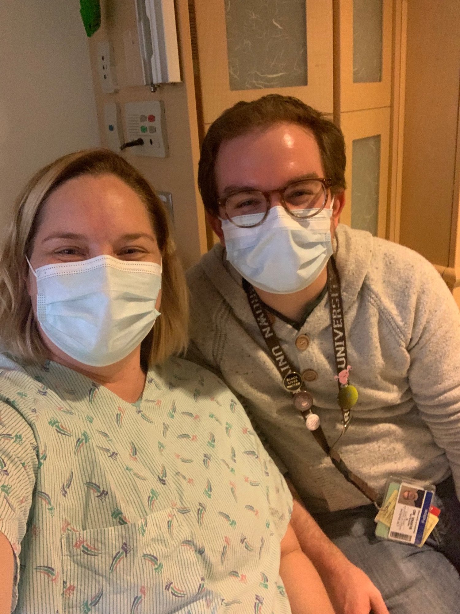
PLOT TWIST!  The patient was Laura, and the MFM was Nick!

IVIG Infusions worked once we got them going! ;)


The ARRIVE Trial

Here’s the RoshReview Question of the Week!

A 32-year-old G1P0 woman at 39 weeks gestation is admitted for induction of labor. Her induction is started with vaginal misoprostol. Cervical ripening by this method is caused by the action of which of the following substances?

Check out the links above to see if you got this week’s question correct!


Actual Title: Labor Induction vs. Expectant Management in Low-Risk Nulliparous Women 

ARRIVE = A Randomized tRial of Induction Verses Expectant Management

Background: 

  • Who did the study? 

    • As many big studies in Ob/Gyn, it was done by the MFMU and the Eunice Kennedy Shriver National Institute of Child Health and Human Development 

    • First author was Dr. William Grobman from Northwestern University (now at Ohio State!)

    • The study was done in 41 hospitals participating in the MFMU 

  • Where was the study published? 

    • Published in NEJM in August 2018 

  • Why was the study done? 

    • In previous observational trials, there was worse perinatal outcomes when delivery before 39w0d was done without medical indication than when there was delivery at full term 

    • However, there was also thought that delivery after 41 weeks can lead to increased perinatal risks (ie. increasing risk of stillbirth) 

    • There was also thought that induction of labor should be avoided if there was no reason to induce  (ie. elective induction) because induction led to increased cesarean section and possible adverse maternal outcomes 

    • There was one previous study in the UK of 619 women 35 years and older that showed increased risk of c-section 

  • What was the objective? 

    • Test if elective induction at 39 weeks would result in lower risk of composite outcome of perinatal death or severe neonatal complication than expectant management among low-risk nulliparous women 

    • So key things: 

      • Purpose of the trial was to look at NEONATAL outcomes (not mom!) 

      • Second: the population they studied was low-risk nulliparous patients, not everyone! 

Methods

  • Who was in the study? 

    • As we said above, the study was done at 41 centers in the United States that were part of the MFMU (ie. most were large academic centers) 

    • Low-risk nulliparous patients were included in the study 

      • Low risk = no maternal or fetal indication to be delivered before 40w5d (ie. hypertensive disorders, fetal growth restriction etc) 

    • They had to be 34w0d-38w6d at the time of enrollment 

      • Patient had to be certain of LMP or if dating was done with ultrasound before 21w0d 

    • Had to have live, singleton fetus in vertex presentation with no contraindication to vaginal delivery and no C/S planned 

    • Patients who were consented to participate were assessed again between 38w0d and 38w6d to ensure they did not have new indication for delivery that would make them ineligible 

      • Patients who were in labor or who had PROM or bleeding were not eligible 

  • How was the study done?

    • Patients who were eligible were randomized in 1:1 ratio to either:

      • Labor induction - assigned to undergo induction of labor at 39w0d-39w4d 

      • Expectant management - had to forego elective induction before 40w5d, and had to have delivery initiated no later than 42w2d 

      • Of note: no specific induction protocol for either group 

    • Randomization was stratified to site 

    • Participants were then followed and data was abstracted from their chart

    • Patients also had interview to rate their labor pain on Likert Scale and also rate their experience with Labor Agentry Scale 

  • What outcomes did they look for?

    • Primary outcome: composite of perinatal death or severe neonatal complications

      • Consisted of one or more of many things (don’t have to list all): perinatal death, need for respiratory support within 72 hours after birth, Apgar of 3 or less at 5 min, HIE, seizure, infection, meconium aspiration syndrome, birth trauma, intracranial or subgaleal hemorrhage, or hypotension requiring vasopressor support 

    • Main secondary outcome: cesarean section 

      • Lots of other neonatal secondary outcomes and maternal secondary outcomes that we don’t need to list 

    • Other prespecified subgroups: race, age >/= 35 or <35, BMI, modified Bishop score at time of randomization of <5 vs. 5 or more 

Results 

  • Participants 

    • Recruited from March 2014 - August 2017

    • Out of 22,533 eligible women, 6106 (27%) consented and were randomized

      • 3062 in induction and 3044 to expectant management 

      • 63% had unfavorable Bishops (<5) 

      • Both groups were similar  

    • 3 in induction group and 7 in expectant management group were lost to follow-up 

    • 6% of induction group and 4.6% of expectant management group had protocol violation 

  • Outcomes 

    • Those in induction group had shorter median time from randomization to delivery than in expectant management group (7 vs. 12 days) 

    • Women in induction group underwent delivery at a significantly earlier median gestational age (39.3 wks IQR 39.1-39.6 vs. 40.0 IQR 39.3-40.7 weeks) 

    • Primary outcome 

      • Occured in 4.3% of neonates in induction group vs. 5.4% in the expectant management group (RR 0.8, 95% CI 0.64-1.00) 

      • Did not change after adjustment for previous pregnancy loss 

      • Neonates in induction group had shorter duration of respiratory support and total hospital stay 

        • Other secondary outcomes for neonates were the same 

    • Secondary outcomes for mom 

      • Cesarean delivery was 18.6% in IOL group vs. 22.2% in expectant management group (RR 0.84, 95% CI 0.76-0.93), p<0.001 

      • HTN disorders of pregnancy was 9.1% in IOL group vs 14.1% in expectant management group (RR 0.64, 95% CI 0.56-0.74), p<0.001 

      • Of note, interestingly there were also higher scores on LAS both immediately after and 4-8 weeks after delivery in the IOL group 

      • Median labor pain was also reported as less (8 vs. 9) in IOL group vs. expectant management group 

        • Of note though, for LAS and median labor pain score, the scores were statistically significant but overall difference was small 

      • Women in the IOL group spent more time on the labor and delivery unity but their postpartum stay was shorter 

      • Subgroup analyses showed no significant difference between group differences 

Discussion 

  • What happened after the study? 

    • Don’t know about your hospitals, but we have begun offering 39 week inductions to all nulliparous patients if they desire them 

      • ACOG also made a statement that reasonable to offer 39 week induction as long as we also take patient preference into consideration 

    • It is not “recommended” but offered 

    • Why? 

      • Some people take this to mean that by having IOL at 39 weeks that we are not only not changing neonatal outcomes, we are also decreasing CS rates and HTN disorders of pregnancy per this study 

      • They also would argue that there is no difference in LAS or pain overall (and if anything, patients feel more agentry and less pain) with IOL

    • What’s the other side of the story? 

      • In 2018, the ACNM responded to the ARRIVE Trial study results 

      • Discussed that potentially by increasing IOLs, we are also increasing the use of hospital resources (ie. staff, capacity of hospital beds, etc) 

      • Also stated that the study criteria were very strict (low risk, nulliparous), and discussed that we should be careful of broadening the outcomes and applying IOL to all patients 

      • Basically: concern that we will be offering IOL to everyone without knowing the actual implications 

    • More to the story 

      • There was a lot of concern about cost and hospital resources , so in 2020 Einerson et al came out with a study looking at cost 

      • Reviewed health-system cost of elective IOL at 39 weeks vs. expectant management in Utah hospitals 

        • No cost difference between expectant vs. IOL 

          • Maternal outpatient antenatal cost were 47% lower in the induction arm, and intrapartum and delivery costs were 16.9% higher 

  • How do we practice now? 

    • One study did look at rates of IOL pre and post ARRIVE 

      • Gilroy et al looked at rates of IOL in the country in patients who were nulliparous who started prenatal care by 12 weeks and delivered at 39 weeks or later  

      • There was a significant increase in IOL after ARRIVE 

        • 36.1% vs 30.2%, OR 1.36

        • Also more likely to deliver by 39w6d (42.8% vs. 39.9%) 

        • Less likely to have a CS (27.3% vs. 27.9%) ← but that is a much higher rate than 18% in ARRIVE 

The Twin Birth Study

Here’s the RoshReview Question of the Week:

Which of the following needs to be met to undergo vaginal delivery with a monochorionic-diamniotic twin pregnancy with vertex twin A?

Check your answer and get a special RoshReview deal for listeners at the links above!


Actual title: A Randomized Trial of Planned Cesarean or Vaginal Delivery for Twin Pregnancy

https://www.nejm.org/doi/full/10.1056/nejmoa1214939 

Background:

  • Where was the study published?

    • NEJM, October 3, 2013

  • Why was the study done?

    • Tthrough the 1990s and 2000s there was a significant rise in twin births in the USA, likely attributed to advancing maternal age (when twinning is more common spontaneously) and the use of reproductive technology – ovulation induction and IVF.

    • In the wake of the Term Breech trial, as well as some observational studies looking at twins specifically, there was concern that breech birth risks could be extended to twins – and practice was changing!

      • In 1995, 53.9% of twin births were by CS. By 2008, this number was 75%. 

    • Not all observational studies were in agreement about the risk of “breech extraction” of a second twin, specifically – so a new study was planned and performed.

  • Who performed the study?

    • The “Twin Birth Study Collaborative Group” – a large multinational collaborative, but with the main site at the University of Toronto and funded by the Canadian Institutes of Health Research – the same funders that brought you the Term Breech Trial!

      • You’ll note a lot of similarities (but also some important differences!) between this study and the Term Breech Trial. We definitely recommend a compare-contrast session!

  • What was the research objective?

    • To compare the risk of fetal/neonatal death or serious morbidity between planned cesarean or planned vaginal delivery for twin pregnancies between 32w0d and 38w6d, if the presenting twin was in cephalic presentation. 

Methods:

  • Who participated and when?

    • Recruitment between December 13, 2003 and April 4, 2011 at 106 centers in 25 countries.

    • Enrolled 1392 patients in the planned cesarean group and 1392 patients in the planned vaginal delivery group.

  • Eligibility:

    • Needed to have:

      • Twin pregnancy between 32w and 38w6d

      • First twin in cephalic presentation

      • Both fetuses alive with EFW between 1500g and 4000g, confirmed by ultrasound within 7 days before randomization

    • Exclusions:

      • Monoamniotic twins

      • Lethal fetal anomalies

      • Other contraindication to labor or vaginal delivery (including 2nd twin being “substantially larger” than the first)

      • Prior cesarean with vertical incision or more than one LTCS

  • Management:

    • Delivery by cesarean or by labor induction was planned between 37w5d and 38w6d

    • If in the CD group, if the first twin delivered vaginally, then a c-section was attempted for the second twin if logistically possible.

    • In the VD group:

      • Continuous EFM was “recommended” during active labor

      • Use of oxytocin and epidural analgesia were left to OB provider discretion

      • After delivery of first twin, use of US was “encouraged” to check second twin presentation

        • If cephalic, amniotomy was delayed until head was engaged and SVD anticipated, unless for other OB indication

        • If non-cephalic, OB decided on best delivery option – spontaneous or assisted breech delivery, total breech extraction +/- internal podalic version, ECV and vaginal cephalic delivery, or intrapartum CD

      • Deliveries were attended by qualified OB experienced in twin delivery, defined as a OB who judged themselves to be experienced at twin delivery and whose department head agreed with this judgment (similarly to Term Breech Trial).

  • Outcomes:

    • Primary: fetal/neonatal mortality or serious neonatal morbidity, assessed up to 28 days after birth.

      • Morbidities included many of the same things in the Term Breech Trial, and were serious neonatal morbidities (for the sake of brevity, we won’t list them out).

    • Secondary: maternal death or serious maternal morbidity, assessed up to 28 days after delivery.

      • Again, this was very similar to the Term Breech Trial. 

    • A number of subgroup analyses were planned for the primary outcome, including by nulliparity; gestational age at randomization; maternal age; presentation of the second twin; chorionicity; and the perinatal mortality rate in the mother’s country of residence. 

Results

  • Who was recruited?

    • Outcome data was available for 1392 women (2783 fetuses/infants) in the cesarean group and 1392 women (2782 fetuses/infants) in the vaginal delivery group. 

    • Baseline characteristics were overall similar, and most patients (82.4%) underwent randomization between 32w0d and 36w6d. 

      • More than half of the infants in each group were born at 37w0d or later. 

        • Around 5-6% in each group were between 32w and 33w6d, and another 42% between 34w0d to 36w6d. 

      • The time from randomization to delivery was similar but slightly different between groups (12.4 vs 13.3 days).

  • In the planned CD group: 

    • 90% had CD

    • 1% had a combined vaginal-cesarean delivery, and 

    • 9% had both twins vaginally.

      • Almost 60% of the CDs were performed before the onset of labor.

  • In the planned VD group:

    • 56% delivered both twins vaginally, 

    • 4% had a combined vaginal-cesarean delivery, and 

    • 40% had a cesarean for both twins.

      • Of those in the VD group who had a CD, 67.5% of them were performed during labor (or another way to look at it, 32.5% had a CD prior to labor in the planned VD group).

    • 95% had an experienced OB present, according to the study definition

  • Primary Outcome:

    • The frequency of composite primary outcome did not differ between planned CD (60, or 2.2%) and planned VD (52, or 1.9%) groups.

      • The only variable that appeared to modify the risk of the primary outcome was earlier gestational age at randomization. 

      • The number of deaths in each group was 24 (0.9%) in CD group and 17 (0.6%) in VD group. 

        • 11 of these deaths in the CD group and 8 in the VD group were before labor onset.

    • In subgroup analyses, there was no significant interaction with the primary outcome with respect to parity, gestational age at randomization, presentation of the second twin, chorionicity, or national perinatal mortality rate. 

    • The second twin was more likely than the first to have the primary outcome, but this was not different between the groups. 

  • Secondary outcome:

    • There were no differences in primary maternal composite outcome rates (7.3% CD, 8.5% VD). 

Impact

  • What is the impact of all of this, and what are we doing now?

    • This paper certainly helped to encourage the training and planning of vaginal delivery of the second twin, including by breech delivery by stating that no increased risk was seen with a policy of planned vaginal delivery. 

      • In ACOG PB 231 on multifetal gestation, it notes that vaginal delivery of a non-cephalic second twin is reasonable, provided an OB with experience is present.

      • That’s key – it’s apparent in this paper that, compared with the Term Breech Trial, there was more emphasis on patient counseling / selection (i.e., 13 day median from randomization to delivery, protocolized assessment of EFW by US within 7 days, 95% presence of “experienced OB”). 

        • And this is heavily noted in the conclusions of the paper – stating “only centers that can provide OB management as specified by the protocol, including ability to perform a CD within 30 minutes if necessary” should undertake this.

  • Methodologically, this group responded to many criticisms of the Term Breech Trial:

    • An improved randomization scheme that was block-based, stratified by gestational age and parity.

    • Improved use of ultrasound and CTG in labor, as well as higher standard of care at all sites to prevent misappropriation of primary outcome.

    • More explicit counseling – happening weeks before delivery on average, rather than in labor!

  • And finally - and most importantly - this represents a well-selected, high-resource, best-case scenario work.

    • For our US listeners who mostly practice in centers where there is ability to perform cesarean within 30 minutes, the Twin Birth Study included:

      • Twins delivering between 32w0d and 38w6d

      • With EFW estimated by US within 7 days of delivery, ranging from 1500g - 4000g

        • Second twin not significantly larger (with expert opinion putting this around a max of 15% discordance)

      • Ability to perform CD within 30 minutes, and use CTG and intrapartum US

      • With someone with experience and ability to perform breech extraction and internal podalic version available