Abortion: Telemedicine and Self-Management

Today we’re joined by Dr. Sarah Gutman, who is an assistant professor of OB/GYN at the University of Pennsylvania, and a recent graduate of their fellowship in complex family planning. She’s joining us today to talk about some of the most important and interesting topics trending in the weeks after the Dobbs vs Jackson Women’s Health: self-managed abortion and telemedicine abortion.

What is telemedicine abortion?

  • Provision of medication abortion care using telemedicine services, typically fully remote but can involve some degree of in-person contact for part of the process, under the supervision of a medical provider.

  • Who are the appropriate candidates for telemedicine abortion?

    • Eligibility criteria for studies evaluating telemedicine abortion have typically included:

      • Pregnancy less than 10 weeks gestation,

      • No contraindications to mifepristone or misoprostol and

      • The ability to receive mife/miso by mail

  • What are the steps of a typical telemedicine abortion visit?

    • Initial consult – confirmation of dating, review of medical history/risks factors, discussion of how medication should be used and expectations for abortion process.

    • Patients should be certain of their LMP within one week, and it should be <77days before anticipated start of mifepristone

    • Evaluate for symptoms or risk factors for ectopic pregnancy, including vaginal bleeding, pelvic pain, prior ectopic, current IUD use, prior tubal surgery

      • Interestingly, the rate of ectopic pregnancy among patients seeking abortion is lower than the general population – between 1.5 – 6 per 1,000 pregnancies compared to about 20 per 1,000 pregnancies in the general population

    • Ensure no contraindications for medication abortion

      • RH type and hemogloblin are not needed

    • Receipt of medications – due to restrictions in mifepristone accessibility, typically this has been through the mail

  • Medication abortion has been covered by the podcast in the past, but as a reminder: the two medications used for medication abortion are mifepristone and misoprostol.

    • Mifepristone is a taken orally as a one-time 200mg dose

    • Misoprostol can be used vaginally, sublingually, or buccally, 800mcg are given initially with the option to repeat a dose if needed.

      • Patients are informed to take within 48h of mifepristone administration.

      • Consider a second dose if GA >63 days or no bleeding in 24 hours.

    • Analgesics, antiemetics – many providers give ibuprofen and Zofran

  • When to seek help:

    • Heavy bleeding soaking >2 pads/hour for more than 2 hours,

    • Passing blood clots larger than a lemon, or

    • Symptoms of blood loss such as feeling dizzy/lightheaded.

  • Follow up

    • Symptoms – can be assessed at 7-14 days through a text, secure messaging, telephone all, or video.

      • Patients are counseled to expect bleeding heavier than a period, and that they may pass blood clots and see some tan/pink tissue.

    • Urine pregnancy tests – given 4 to 6 weeks following the abortion

What is the evidence behind telemedicine abortion?

  • Efficacy is very high – around 95% of abortions are completed without needing a procedure.

  • Complications are exceedingly rare.

    • Around 6% of patients visit an ER or urgent care center related to the abortion

    • The rate of adverse events is less than 1%, with hospitalization <0.5%, transfusion 0.4%, infection <0.1%

What is self-managed abortion?

  • Self-managed abortion has also been referred to as self-sourced medication abortion (SSMA)

  • Society of Family Planning definition:

    • “It refers to any action taken to end a pregnancy outside of the formal healthcare system, and includes self-sourcing mifepristone and/or misoprostol, consuming herbs or botanicals, ingesting toxic substances, and using physical methods.”

  • Historically, people fearing criminalization or unable to access abortion care often turned to unsafe or invasive methods of self-managing their abortion – think of the abortion scene in ‘Dirty Dancing’ and the use of a coat-hanger as a sign of an unsafe abortion.

    • However, increased access to the medications used for abortion, in particular misoprostol, had made self-managed abortion much safer and more effective.

    • Other reasons besides access that people may choose self-managed abortion, including privacy, discomfort with the available medical services, and person safety.

What are the components of SMA?

  • Similar to telemedicine abortion, SMA includes assessment of eligibility, administration of abortion medications, management of the abortion process, and assessment of abortion completion.

    • These actions are all taken without the formal guidance of a healthcare provider.

    • People who self-manage their medication abortions should be able to estimate their gestational age using their last menstrual period and be aware of their cycle regularity and any contraception use.

  • There are many clinical resources available online, including through the Reproductive Health Access Project, Doctors without Borders, and Aid Access.

  • The WHO recommends mifepristone followed by misoprostol.

    • However, if mifepristone is not accessible, misoprostol can be used alone, typically 800 mcg used vaginally, sublingually, or buccally repeated every 3 hours or up to 3 doses until expulsion occurs.

  • How common is SMA?

    • Recent cross-sectional data suggests 7% of individuals in the US attempt SMA at some point in their lifetime, and this is likely growing due to increased restrictions on abortion access.

  • What is the safety and efficacy of SMA?

    • Data is limited: it’s difficult to study something that is outside the healthcare system.

    • However, from the data we have available and by extrapolating data from the telemedicine abortion models with lowest amount of supervision, self-managed abortion using mifepristone and misoprostol appears to be as safe and effective as medication abortion within a clinical setting.

    • A meta-analysis of misoprostol alone regimens used <91 days gestation found a 6.8% ongoing pregnancy rate

      • Serious adverse events occur <1% of the time.

  • How can providers support patients who have chosen self-managed abortion?

    • When people are criminalized for abortion, it is often due to a healthcare provider reporting them to the police.

    • Currently, there are no mandated reporting laws for healthcare providers.

    • There is legal help available for patients concerned about their options and criminalization, such as If/When/How

      • People of color and low-income individuals are most likely to be targeted and disproportionately criminalized.

Summary

  • Telemedicine abortion is the provision of medication abortion through telehealth under a healthcare providers supervision. Self-managed abortion is actions taken outside the formal healthcare setting to end a pregnancy.

  • Both telemedicine abortion and self-managed abortion using mifepristone and misoprostol are remarkably safe and effective.

  • While protocols vary, typically patients receiving telemedicine abortion should be at or below 10 weeks gestation, should not have any risk factors or symptoms concerning for ectopic pregnancy, and should not have any contraindications to taking mifepristone or misoprostol. After taking their medications, they should be able to monitor their vaginal bleeding and cramping and take a home urine pregnancy test in 4-6 weeks to confirm completion of the abortion.

  • Importantly, there are no laws mandating that healthcare providers report patients for suspected self-managed abortion. If patients are concerned about criminalization there are legal resources available such as If/When/How.

Additional Resources

Exercise in Pregnancy

Additional reading for today in ACOG Committee Opinion 804!

What are the definitions of physical activity and exercise? 

  • Per ACOG: 

    • Physical activity: bodily movement produced by contractions of skeletal muscles in all stages of life

    • Exercise: physical activity consisting of planned, structured, and repetitive bodily movements done to improve one or more components of physical fitness 

    • Physical activity can maintain and improve cardiorespiratory fitness, reduce the risk of obesity and associated comorbidities, and results in greater longevity 

Is exercise safe in pregnancy?

  • Yes!

    •  Not just in pregnancy, but for all individuals, the US Department of Health and Human Services Physical Activity Guidelines for Americans says: 

      • At least 150 min of mod intensity aerobic activity per week

      • Recommended also in pregnancy and postpartum period 

      • Those that engaged in vigorous-intensity aerobic activity or who were physically active before pregnancy can continue those activities 

      • Few maternal conditions that would not allow aerobic exercise 

  • What are the downsides of not exercising?

    • Physical inactivity is the 4th leading risk factor for early mortality worldwide 

    • Physical inactivity and weight gain in pregnancy have been recognized as independent risk factors for maternal obesity and pregnancy related complications like GDM  

  • What are the benefits of exercising? 

    • Increased likelihood of vaginal delivery! 

    • Low incidence of: excessive weight gain, GDM, gestational hypertensive disorders, preterm birth, c-section, lower birth weight 

  • What exercises are safe? 

    • Aerobic: walking, stationary bike, aerobic exercises, dancing, stretching, water aerobics 

    • Anaerobic: resistance training (weights, elastic bands) 

    • Borg rating of perceived exertion: 6 - 20 (6 to 7 is very, very light, and 19-20 is very, very hard) 

      • Recommend moderate intensity, which is about a 13-14 (somewhat hard) 

        • About 20-30 min per day for most days of the week 

      • Also talk test: if you can talk while exerting yourself, likely not over exerting 

Note: ACOG issued a correction to this table; it should read “First Trimester, Less Than 12 Weeks Gestation.”

What are some modifications we should consider in pregnancy? 

  • Changes in pregnancy 

    • Weight gain, difference in weight distribution 

      • About 60% of pregnant patients will experience low back pain 

    • Increase in blood volume, heart rate, stroke volume, cardiac output = normal (recall our very first few episodes on physiologic changes in preg!)

      • Maintaining supine positioning after 20 weeks may lead to decrease venous return → can lead to SOB, dizziness, hypotension, etc 

    • Minute ventilation increases by 50% 

  • Other modifications: 

    • Remember to stay well hydrated, wear loose-fitting clothing, and avoid high heat and humidity (ie. hot yoga) 

    • Exercise by itself isn’t expected to increase body temp to point of concern 

  • Fetal response 

    • Studies show some minimal to mod increase in fetal heart rate by 10-30 bpm during maternal exercise 

    • Three meta-analyses show that there is minimal to no difference in birth weight 

    • However, women who continue to exercise vigorously in third trimester are more likely to deliver babies weighing 200-400g less than controls, though no increased risk of FGR 

When to stop exercising 

  • Don’t exercise if:

    • Have vaginal bleeding, abdominal pain, regular painful contractions, leaking fluid 

    • Dyspnea before exertion, dizziness, headache, chest pain

    • Muscle weakness affecting balance, calf pain or swelling  

Special considerations 

  • Obesity

    • Encourage patients to have healthy lifestyle modification in pregnancy that include physical activities and judicious diets 

    • Can start with low-intensity, short periods of exercise if not exercising already 

    • Then can build up gradually 

  • Athletes

    • Vigorous-intensity exercise even in 3rd trimester appears to be safe and healthy for most pregnancies 

    • Further research is needed for exercise intensity exceeding 90% of max heart rate  

Perinatal Mental Health, feat. Dr. Tiffany Moore-Simas and Dr. Nancy Byatt

Today on the podcast, we’re addressing perinatal mental health. While we’ve talked about depression on the show before, there’s so much more in this sphere as we’ll discuss today.


Joining us are two experts in this field who share their passion for this work with us. Dr. Tiffany Moore Simas is Chair and Professor of OB/GYN at UMass Memorial Health and UMass Chan Medical School as well as co-Chair of the ACOG Maternal Mental Health Expert Work Group. And Dr. Nancy Byatt is a tenured Professor of Psychiatry and OB/GYN at UMass Memorial Health and UMass Chan Medical School. Both serve as senior leaders with the Massachusetts Perinatal Psychiatry Access Program, MCPAP for Moms, and Lifeline For Moms.


Importance of Perinatal Mental Health

  • Mental health conditions are the most common complications of pregnancy – 1 in 5!

    • More common in adolescents, veterans, marginalized populations (BIPOC, poverty).

  • Untreated mental health conditions carry both short- and long-term consequences that can affect whole family:

    • o   Less engagement in medical care

    • o   Smoking, substance use

    • o   Preterm delivery, low birth weight, NICU admission

    • o   Lactation challenges, bonding issues

      • Parent with untreated mental health disorder is considered an Adverse Childhood Experience (ACE) for the infant.

    • o   Adverse partner relationships

  • Mortality: leading cause of preventable maternal mortality.

    • 100% of maternal deaths due to mental health, including suicide, overdose, are preventable!

  • Underdetected and undertreated

  • OB/GYNs can screen and help manage mental health conditions. The majority (80%) of depression, for example, is managed by primary care providers, not psychiatrists. As obstetric care clinicians, we are the primary care providers to pregnant and postpartum individuals and thus, we should be providing mental health care!

Screening for Perinatal Mood and Anxiety Disorders

  • In this context, perinatal refers to during pregnancy and the first year after pregnancy ends

  • Perinatal Mood and Anxiety Disorders primarily include depression, bipolar disorder, and anxiety or anxiety-related conditions (generalized anxiety disorder, PTSD, OCD).

  • Screens should be performed with validated tools that query the last 7-14 days of symptoms for anxiety and depression.

    • o   Validated tools:

      • PHQ-9, EPDS (depression)

      • GAD-7 (generalized anxiety)

    • o   ACOG recommends screening patients at least once during the perinatal period for depression and anxiety symptoms. If a patient is screened during pregnancy, additional screening should occur during the comprehensive postpartum visit.

      • We recommend screening: new OB visit, later in pregnancy (i.e., 3rd trimester) and postpartum given the almost even distribution of onset predating pregnancy, onset in pregnancy, and onset postpartum.

    • o   Data suggests that early detection and treatment improves outcomes.

  • Bipolar disorder screening:

    • o   In one study, 1 in 5 patients screening positive for postpartum depression actually had bipolar disorder.

      • Recall: bipolar disorder can worsen with antidepressant treatment (unopposed SSRIs) – thus, need to screen for bipolar before initiating pharmacotherapy and ideally universally to prevent harm!

    • o   Patients with bipolar disorder have higher risk of postpartum psychosis

      • Rare: 1-2/1000 perinatal individuals; but 70% have bipolar disorder!

      • 4% risk of infanticide with postpartum psychosis

      • This is a psychiatric emergency.

        • Often occurs within the first days of delivery and most cases occur within the first 3 weeks.

    • o   Screening options:

      • Mood Disorder Question (MDQ) – self administered

      • CIDI – clinician administered with branching logic

    • o   Appropriate to refer to psychiatry if bipolar disorder is suspected – more on resources to help later!

Positive Screening   General Principles of Treatment

  • Just like a glucola, our questionnaires for mental health concerns are screening tests. Subsequent assessment is critical to confirm diagnosis.

    • o   See resources collection at the end of these notes for help!

  • For depression and anxiety, there are three pillars of treatment:

    • o   Psychotherapy

    • o   Pharmacotherapy or medication

      • o   Adjunctive interventions

  • Treat based on level of severity. For information on assessing and treating perinatal mental health conditions, visit the ACOG website.

  • If pharmacotherapy is indicated/started, patients may have some concerns:

    • o   Provide reassurance

    • o   Frame risk/benefit discussion in treated disease vs. untreated disease as not treating is associated with risks - just like any other disease!

    • o   Use lowest effective dose and monotherapy when able

  • Find more information on educating patients about treatment on ACOG’s website.

Concerns for Suicidality or Harm To Baby

  • These can represent urgent clinical scenarios and further assessment and response is critical:

    • o   Thoughts of harming self or baby are common yet not all are necessarily a psychiatric emergency.

    • o   When assessing for risk of harm to self or others it is important to assess:

      • Ideation – Do they have thoughts of harming themselves or someone else?  Are the thoughts fleeting or do they persist?

      • Intent – Are they intending to act on it? Have they thought of how they could do harm themselves or someone else or die by suicide?

      • Plan - Are they planning to act on it?  Have they developed a plan for how to die by suicide or to harm someone else?

    • o   If you are concerned that the patient is at risk of harm to self or others, then it is important to obtain further assessment which includes an evaluation for whether the patient may need psychiatric hospitalization

    • o   Regardless of whether these are a psychiatric emergency, the presence of thoughts of harming self or baby are indicative of higher illness severity.

  • More information on ACOG’s website.

 Resources for Integrating Perinatal Mental Health Care into Your Practice


Sickle Cell Disease, feat Dr. David Abel

Here’s the RoshReview Question of the Week:

Which of the following is a precipitating factor for a painful crisis in a pregnant woman with sickle cell disease?

Check out the answer at the links above and get a sweet deal on RoshReview!


Today we’re joined once again by Dr. David Abel, an assistant professor in the Department of Obstetrics and Gynecology at Oregon Health Sciences University. Dr. Abel has previously joined us to talk about thalassemias and von Willebrand’s disease — he shares his passion for blood disorders again with us today to talk more on sickle cell!

Listen to our last sickle podcast here. 

Epidemiology of Sickle Cell Disease (HbSS)

  • Most common inherited hemoglobinopathy in the United States, and in fact is the most common inherited disease worldwide.

    • Autosomal recessive fashion — both parents need to be carriers.

  • Affects approximately 10 million people worldwide and approximately 100,000 people in the United States.

    • This translates into a prevalence of about 1 in 375 who have the disease.

  • Predominantly affects people of African ancestry

    • Two thirds of those that are affected reside in West Africa.

    • 50% of children born with sickle cell disease are born in Nigeria.

  • Sickle Cell Trait: 1 in 12 are carriers.

    • In the United States, approximately 7% to 9% of the African American population.

    • As the gene is widely distributed, other populations may be affected including those residing in the areas of the Mediterranean, Caribbean, South and Central America, as well as East India.

  • More than 90% of children with sickle cell disease in the United States survive into adulthood.

    • Compared to the general population, however, their lifespans are two or threedecades shorter and limited by both acute and chronic morbidity.

  • Sickle cell trait confers a survival advantage in malaria-endemic regions such as in sub-Saharan Africa where almost 80% of individuals with sickle cell anemia live — resulting in “positive screening” with respect to human evolution.

Hemoglobin Structure in Sickle Cell and Pathophysiology

  • Remember: thalassemias represent quantitative defects globin synthesis. By contrast, hemoglobin S is characterized by a qualitative defect of the beta globin gene.

    • HbS results from a single nucleotide substitution, an adenine- to-thymine substitution in the sixth codon of the beta globin polypeptide which replaces glutamic acid with valine.

      • From this one amnio acid change, rather than forming tetramers, under conditions of low oxygen tension, this hemoglobin S forms long inflexible chains or fibers.

      • They distort the red blood cell membrane, resulting in this sickled shape.

      • These distorted red blood cells are destroyed by the reticuloendothelial system, resulting in a moderate to severe anemia.

        • Compared to the normal life span of a red blood cell of 120 days, the life span of these sickled red blood cells is reduced to an average of 15 days.

  • These distorted red blood cells clog up the microvasculature —> obstruction and local ischemia which clinically manifests as a vasoocclusive crisis.

    • Repeated vasoocclusive crises can lead to interruption of normal perfusion of multiple organs, including the spleen, lungs, kidneys, heartand brain.

    • Adults with sickle cell disease are essentially functionally asplenic — increased incidence and severity of infection in patients with sickle cell disease.

  • Sickled red cells are also prone to lysis which releases free hemoglobin.

    • Damages the endothelium and may also lead to thrombosis.

    • Also consumes nitric oxide, an important vasodilator and thus can lead to an exacerbation of the ischemia.  

Testing for Sickle Cell Disease and Trait

  • Foundation of screening: CBC, Hb electrophoresis. High suspicion in patients with family history / ancestry and MCV < 80.

    • HbSS (disease) electrophoresis:

      • 85-95% hemoglobin S

      • Remaining mostly hemoglobin F, small component of hemoglobin A2

    • HbSB (trait) electrophoresis (assuming normal second beta gene):

      • 50-60% hemoglobin A (normal adult Hb)

      • 35-45% hemoglobin S

      • Small amounts hemoglobin F, hemoglobin A2

  • There are many other sickle genotypes:

    • Homozygous hemoglobin SS constitutes about 70% of these genotypes.

    • Hemoglobin C, which differs from hemoglobin S only in that the amnio acid lysine instead of a valine replaces glutamic acid in the beta globin gene, can exist in combination with hemoglobin S, thus is called hemoglobin SC disease.

    • Hemoglobin S may coexist with beta-thalassemia.

      • Hemoglobin S beta thalassemia zero is also identified as sickle cell anemia and is just as severe as hemoglobin SS.

      • Hemoglobin S beta thalassemia plus is not as severe as there is some hemoglobin A that is preserved.

Maternal and Fetal Considerations with Sickle Cell

  • During pregnancy, the increase in red blood cell mass that normally occurs does not in those with sickle cell anemia.

    • 50%–70% of pregnancies with sickle cell disease require at least one hospitalization

    • 30%–40% will require a transfusion.

    • In the United States, the maternal mortality rate is approximately 10 times higher than it is for patients without sickle cell disease.

  • Vasoocclusive Crisis / Pain Crisis is most common cause of recurrent morbidity.

    • Can be precipitated by such factors as cold, physical exertion, dehydration and stress.

    • Opioids are a mainstay of treatment for a pain crisis — it is important not to withhold treatment for these patients.

  • Acute chest syndrome severe life-threatening form of a vasoocclusive crisis

    • Presents similarly to pneumonia.

      • Fever, tachypnea, chest pain, hypoxia and infiltrates noted on chest x-ray.

      • In addition to infectious agents, acute chest syndrome may also result from fat emboli, intrapulmonary aggregates of sickled red blood cells, atelectasis or pulmonary edema.

    • Patients with a history of frequent hospitalizations and/or episodes of acute chest syndrome correlate with increased risks during pregnancy.

    • The treatment of acute chest syndrome typically consists of antibiotics, usually ceftriaxone and azithromycin, pain control, and if needed oxygen and transfusion.

  • Other complications

    • Stroke: occurs in almost 25% by the age of 25

    • Splenic sequestration / asplenia

    • Acute renal failure

    • Acute cholecystitis

    • Pulmonary hypertension: 6 to 11% of patients with sickle cell disease

    • Venous thromboembolism: ocurs in 10-25% of those with sickle cell disease by age 40

      • In pregnancy risk elevates: 2x increased risk of stroke, 5x increased risk of cerebral vein thrombosis, 2x increased risk of pulmonary embolism, 2.5x increased risk of deep vein thrombosis.

    • Maternal infection complications: asymptomatic bacteriuria, pyelonephritis, sepsis and an almost ten-fold increased risk of pneumonia.

  • Placental Consequences:

    • Placental hypoperfusion with endothelial damage is the main contributor to adverse pregnancy outcomes.

    • Increased risk of preeclampsia and eclampsia, placental abruption, antepartum bleeding and alloimmunization.

  • Fetal consequences:

  • 2x increased risk of preterm birth

  • 3x risk of small-for-gestational age

  • 4x increased risk of stillbirth.

    • Serial fetal growth assessments and antepartum testing are needed.

  • Possible increased risk of neonatal abstinence syndrome due to the use of opioids to treat pain crises.

Should transfusion be used prophylactically?

  • 2015 meta-analysis of 12 observational studies with almost 1300 patients demonstrated a reduction in both maternal and perinatal mortality as well as a reduction in pain events and preterm birth.

  • 2016 Cochrane review that included only randomized controlled trials did not demonstrate a benefit with prophylactic when compared with selective transfusion.

  • Management strategy:

    • CBCs should be checked frequently, and a goal of maintaining a hemoglobin around 10 (same might say up to 12) and a percentage of hemoglobin S less than 35 to 40% is reasonable.

    • It is important to avoid iron overload when considering transfusion therapy which may lower target Hb for some individuals.

Treating Sickle Pain Beyond Opioids

  • Amitriptyline

  • Gabapentin

  • Selective serotonin reuptake inhibitors (SSRIs) and selective norepinephrine reuptake inhibitors (SNRIs)

  • Complementary (CAM) therapies

  • Hydroxyurea

    • Mainstay of treatment in the non-pregnant patient, as it reduces the risk of a vasoocclusive crisis and acute chest syndrome, thereby leading to improved survival and quality of life.

      • Patients with sickle cell disease who have higher amount of fetal hemoglobin tend to do better, and hydroxyurea increases fetal hemoglobin as well as reduces red blood cell adhesion and increases nitric oxide, the vasodilator we discussed earlier.

    • Has been found to cause birth defects in animals, although it has not been found to increase the risk of birth defects in humans. Still, it is generally avoided during pregnancy.

 Preconception Counseling for Sickle Cell

  • Discuss the increased risk of both maternal and fetal complications, though many can have a successful pregnancy.

    • Know history — past need for transfusions, the frequency of hospitalizations due to vasoocclusive crises and if there is a history of acute chest syndrome.

  • Due to the possibility of iron overload due to multiple transfusions, it is important to check a ferritin prior to prescribing iron.

  • ACOG recommends 4 mg of folic acid daily.

  • Screen for hypertension and treat if warranted to maintain a blood pressure less than 140/90.

  • A dilated eye examination performed by an ophthalmologist should be performed if not done within the past year.

  • If there are any concerns for nephropathy, refer to nephrology — screen for proteinuria.

  • If there are any concerns for pulmonary hypertension, an echocardiogram is recommended with cardiology referral if needed.

  • As most patients are functionally asplenic, pneumococcus, haemophilus influenza type b, and meningococcus immunizations are recommended.

  • Check antibody screen — possible risk of alloimmunization.

Cure for Sickle Cell Disease?

  • Have been accomplished with hematopoietic stem cell transplantation.

    • In this case, the donor may be related or unrelated.

  • Stem cell transplantation offers a cure, but can result in death, graft rejection, graft versus host disease, and sterility due to chemotherapy.

  • Gene therapy for sickle cell disease, where patients receive their own genetically modified hematopoietic stem cells, is still experimental and there are several clinical trials underway.  

Final Important Points on Managing Sickle Disease in Pregnancy

  • Hydroxyurea should be discontinued if it has not been already.

    • There are some who might consider restarting this after the fetal anatomical survey shows no evidence of abnormalities.

  • Chelation agents should also be discontinued.

  • Low-dose aspirin for preeclampsia risk reduction is recommended.

  • Monthly urinary cultures monitoring for asymptomatic bacteriuria and watching for any signs or symptoms of pyelonephritis.

    • Also reasonable to do in patients with sickle trait as risk is also increased in that population.

  • Frequent CBC monitoring, usually monthly is reasonable.

  • Interval fetal growth assessments every 3-4 weeks are recommended.

  • Antepartum testing starting at 32 weeks is indicated, with a delivery goal of 37 to 39 weeks.

  • Watch for preeclampsia is very important.

  • If a cesarean delivery is required, a preoperative transfusion may be prudent to increase hemoglobin levels to 8 to 10 g/dl.

  • Thromboprophyalxis: SCDs definitely; anticoagulation should be individualized.

    • Assuming the patient did not have a thromboembolic event during the pregnancy, could consider prophylactic low-molecular weight heparin for six weeks postpartum.

Dermatoses of Pregnancy, feat. Dr. Laura Hanks

Here’s the RoshReview Question of the Week!

A 30-year-old G1 woman presents at 34 weeks gestation to the office with severe itching and a rash that suddenly developed across her abdomen starting around her umbilicus. The rash is mostly urticarial papules and plaques, but there are a few scattered bullae. Which of the following is required to confirm the suspected diagnosis?

Check your answer at the links above and get a special deal on RoshReview!


Today we welcome to the podcast Dr. Laura Hanks, who is an Assistant Professor in the Dept. of OB/GYN at the University of Wisconsin - Madison. She was previously in private practice in Olympia, WA, and did her residency training at the University of Rochester in New York.

Dermatoses are a pretty confusing topic — so if you have access to some textbooks through your medical school or residency libraries, Dr. Hanks bookmarked a few good chapters:

  • Gabbe’s Obstetrics. Normal and Problem Pregnancies. 8th edition. Chapter 56: Skin disease in pregnancy.  

  • Habif’s Clinical Dermatology. Chapter 6: Urticaria, Angioedema and Pruritus. 

  • Creasy and Resnik’s Maternal-Fetal Medicine. 8th edition. Chapter 69: The Skin and Pregnancy. 

What are dermatoses?

  • Dermatoses of pregnancy refers to a group of skin diseases encountered predominantly during pregnancy or immediately postpartum.

  • In general there is a lack of understanding of the pathogenesis of most of these conditions and therefore a lack of specific diagnostic criteria.

In the podcast, we use a case: 36y G1 at 22 weeks who develops severe pruritus of abdomen, spreading to thighs.

Differential Dx of Dermatoses

  • Pruritis of pregnancy (pruritis gravidarum)

    • Reported in 1.5-2% of pregnancies and occurs most frequently over the abdomen.

    • Usually just pruritus and no rash

    • Often presents in the 3rd trimester

    • Reversible form of hormonally triggered cholestasis

    • Runs in families and typically recurs in subsequent pregnancies

    • Severe pruritus with no primary skin lesions

    • Pruritus on palms and soles that later becomes more generalized

    • Itching correlates with elevated serum bile acid levels and sometimes aminotransferases

    • Bile acids can pass into fetal circulation and cause placental anoxia and cardiac depression which can preclude premature birth, stillbirth, neonatal respiratory distress syndrome, vitamin K deficiency and coagulopathy in the other and newborn.

  • Polymorphic eruption of pregnancy (PEP) or pruritic urticarial papules and plaques of pregnancy (PUPPPs)

    • Affects 1 in 130 to 1 in 300 (0.6%) in the US.

    • This often presents in the 3rd trimester or postpartum with resolution during the postpartum period more commonly in primigravidae pts

    • Abdominal distension causes damage to the connective tissue that in turn triggers an inflammatory response.

    • Associated with multiple gestation due to higher levels of progesterone, which has been shown to aggravate the inflammatory process at the tissue level.

    • Intensely pruritic urticarial rash with erythematous edematous papules and plaques that starts in the abdominal striae and spares the umbilicus. Can include urticarial and sometimes vesicular, purpuric, or targetoid lesions similar to PG or erythema multiforme. Lesions usually spare the palms and soles.

    • This is a clinical diagnosis with no lab findings and no indication for biopsies. If a biopsy is preformed it will often show a nonspecific perivascular lymphohistiocytic infiltrate +/- eosinophils

    • There are also no known risks to the fetus.

  • Impetigo herpetiformis or pustular psoriasis of pregnancy

    • Often associated with reduced calcium or Vitamin D

    • Eruption usually during 3rd trimester, most cases resolve postpartum

    • Characterized by numerous grouped, discrete, sterile pustules at the periphery of erythematous patches

    • Lesions typically originate on flexures and progress to trunk. Spares face, palms and soles.

    • Constitutional symptoms can be common including fever, malaise, diarrhea and vomiting with dehydration.

    • Lab findings include a luekocytosis, elevated erythrocyte sedimentation rate, hypocalcemia and decreased vitamin D levels

    • Risk of stillbirth and fetal abnormalities secondary to placental insufficiency

    • Maternal prognosis is very good with early diagnosis and aggressive treatment, however the increased risk of perinatal mortality may persist despite maternal treatment

    • Diagnosis is based on histopathology that shows typical features of pustular psoriasis. Direct and indirect skin immunofluorescence is negative

  • Pemphigoid gestationis

    • Unfortunately sometimes referred to has herpes gestationis, however it is not related to infection by herpesvirus. This synonym was used to refer to the grouped (herpetiform) nature of the blisters, which often are not herpetiform.

      • It is best to avoid the term herpes gestationis because of the risk for misleading patients and misinformed health care workers; not using the term avoids potentially inappropriate treatments for herpesvirus.

    • Rate in the US is 1 in 50,000 (0.002%)

    • Often presents in 2nd or 3rd trimester and sometimes postpartum (25%) with extremely pruritic, urticarial lesions that typically begin on the abdomen and trunk, that commonly involve the umbilicus. These urticarial plaques can then very quickly progress to widespread bullous lesions that may affect palms of hands and soles of feet. There is often a flare at the time of delivery with resolution during the postpartum period.

    • Lesions can be similar to PUPPs, however PUPPs lesions begin in abdominal striae and spares the umbilicus unlike PG.

    • Suggested pathogenesis is complement-fixing IgG antibodies and complement C3 react with amniotic epithelium of placental tissues and basement membrane of the skin causing an autoimmune response resulting in tissue damage and blister formation.

    • Definitive diagnosis is based on biopsies of the lesions that will show skin direct immunofluorescence shows linear deposition of IgG and C3 along basement membrane.

      • PG recognizes the same antigen as bullous pemphigoid and they do share certain features; however PG itself is confined only to pregnant women and women affected by gestational trophoblastic disease.

    • Skin histopathology shows a spongiotic epidermis and marked papillary derma edema and an eosinophilic infiltrate

    • There is an association between PG and Graves, therefore if you have a pt with PG that is an indication to check thyroid function tests

    • Given the increased risk of small for gestational age infants and preterm delivery, it is recommended to monitor growth US after diagnosis.

    • Unfortunately, there are risks to the fetus which include being born with lesions that are transient due to passive transfer of IgG1 antibodies, increased risk of SGA, preterm birth and IUFD.

  • Atopic eruption of pregnancy

    • This accounts for over 50% of pruritic dermatoses in pregnancy.

    • Most likely presents in the 1st and 2nd trimester with resolution in the postpartum period. This earlier onset may help distinguish from other dermatoses in pregnancy.

    • Features of patchy eczema and papular/prurigo lesions that are located on flexural surfaces, neck, chest, and trunk

    • Serum IgE is elevated

    • No known risk to the fetus

  • Prurigo of pregnancy

    • In the US this occurs in 1 in 300 to 1 in 450 pregnancies

    • Presents in the 2nd and 3rd trimester

    • Grouped excoriated or crusted papules over the extensor extremities and occasionally the abdomen

    • There are no laboratory findings. There may be elevated IgE levels on serologic tests. Previous reports of unfavorable fetal outcomes have not been confirmed.

  • Pruritic folliculitis of pregnancy or follicular papulopustular eruption

    • Rare, exact prevalence is unknown (~30 cases reported). Etiology remains unknown

    • Presents as pruritic follicular erythematous papules and pustules that primarily affect the trunk in the 2nd and 3rd trimester.

    • Biopsy is usually unhelpful, however histopathology is that of folliculitis. Special stains, skin immunofluorescence and serologies are negative.

    • There may be an association with decreased birthweight

  • And don’t forget derm conditions that are not unique to pregnancy!

    • Allergic contact dermatitis

    • Drug reaction

    • Atopic dermatitis or eczema

    • Erythema multiform

    • Scabies

    • Superficial fungal infections

    • Folliculitis

    • Urticaria

    • Vasculitis

    • Secondary syphilis

Habif’s Clinical Dermatology

In the case, we obtain an H&P —

  • She began itching on her upper thighs that then spread to her abdomen, chest, back and arms over several days.

  • She then experienced severe pruritus on the palms of her hands and soles of her feet. She described having to take her shoes off at work and soak them in ice baths and often sleeping with an ice pack between her hands to sooth the itching.

  • Benadryl did not seem to help neither did OTC steroid creams.

  • On exam there were numerous pink-salmon colored annula papules and plaques as well as urticaria with scale within umbilicus, flank, thighs and back. On bilateral medial aspects of feet there were pink-red vesicles with petechial border. 

What next? Dermatology referral and biopsies.

Biopsy results: Positive linear deposition of IgG and C3 antibodies along the basement membrane, suggestive of pemphigoid gestationis!

Treating Dermatoses

  • Generally, same treatment principles apply to all of the specific dermatoses of pregnancy.

    • Milder disease is treated with topical emollients, calamine lotion, cool compresses or baths, and topical corticosteroids.

      • Topical corticosteroids (e.g., hydrocortisone, triamcinolone) are classified as FDA pregnancy category C drugs in the old system, but they are still widely used during pregnancy when the possible benefits outweigh the risks for minimal percutaneous absorption.

  • Intrahepatic cholestasis of pregnancy

    • Ursodeoxycholic acid 15 mg/kg/day daily, BID or TID until delivery

  • PUPPs

    • Topical antipruritic medications, topical steroids and oral antihistamines.

    • In cases of severe pruritis, a short course of oral steroids can be used.

  • Impetigo herpetiformis

    • Systemic steroids are first-line with prednisone dose up to 60-80 mg/day

    • Calcium and vitamin D replacement as needed. Can lead to remission of eruption

  • Pemphigoid gestationis

    • The cornerstone for treatment of Pemphigoid gestationis is oral steroids. Therapy should be directed toward suppressing new lesions and relieving intense pruritus.

    • The majority of patients will respond rapidly to a relatively low-dose of prednisone (20 to 40 mg/day), however the dose may need to be uptitrated according to clinical response as high as 180 mg/day

      • Prednisone should be tapered slowly once new blister formation is suppressed

    • ~75% of patients will experience resolution or at least improvement in the late 3rd trimester, but b/c PG typically flares at delivery, steroid dose can be increased in anticipation of birth.

    • Patients at risk for prolonged or chronic PG are often older with higher parity, more widespread lesions and a history of PG in a prior pregnancy.

  • Atopic eruption of pregnancy

    • Topical steroids, antihistamines, UVB phototherapy

  • Prurigo of Pregnancy

    • Moderately potent topical steroids and oral antihistamines

    • Short course of oral steroids is rarely required

  • Pruritic folliculitis of pregnancy

    • Low-potency or midpotent topical steroids, benzolyl peroxide and UVB.

Ultimately, the patient in our podcast went on to deliver this pregnancy at 38w4d after a cesarean section for fetal indications, without any signs of neonatal blistering (can be seen in 5-10% of babies of mothers with PG!).

The patient ultimately went on to become pregnant a second time, and this time had lesions at 13 weeks. By 25 weeks, prednisone was no longer providing relief!

  • Treating refractory PG

    • A number of alternative treatments have been tested, including:

      • Intravenous immune globulin (IVIG)

      • Plasmapheresis

      • Rituximab

      • Cyclosporine A

      • Azathioprine

      • Dapsone and MTX can sometimes be used postpartum

    • With shared decision making the pt, her MFM and dermatologist elected to proceed with IVIG infusions which were 3 consecutive days every month until the end of pregnancy.

    • Her symptoms improved but never completely resolved during pregnancy and she continued the prednisone along with IVIG. She had a scheduled repeat c-section at 37w3d with stress-dose steroids given at delivery. She gave birth to a healthy male infant weighing 8lbs 1oz without any lesions.

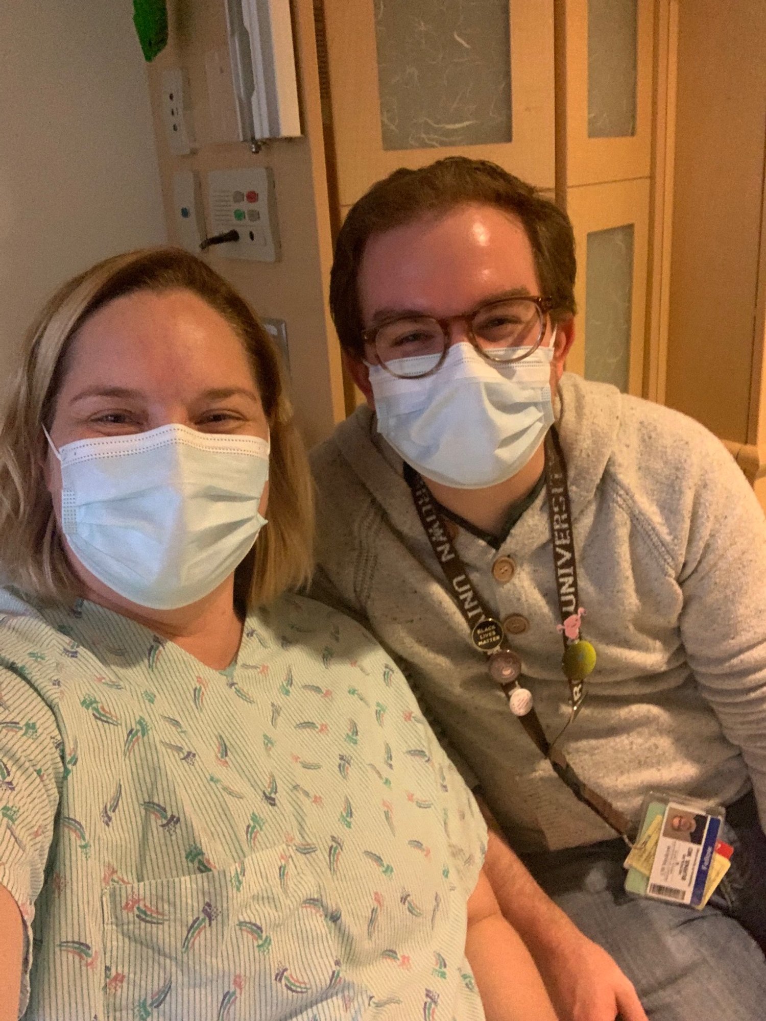
PLOT TWIST!  The patient was Laura, and the MFM was Nick!

IVIG Infusions worked once we got them going! ;)