Adnexal Masses Part IV: Sex Cord Stromal Tumors

Thanks for sticking with us until the end of this adnexal mass journey! Today we’re going to cover some rare tumors that always find themselves on CREOGs — the sex cord stromal tumors. These only comprise about 1.2% of primary ovarian cancers. Most people are fortunately diagnosed at an early stage due to the fact that symptoms tend to be much more overt with these types of tumors.

Granulosa Cell Tumors

There are two subtypes of granulosa cell tumors: adult and juvenile. Adult type comprises 95% of these neoplasms, and generally occur in women aged 50-54 years. Juvenile type typically develops before puberty. It has a higher proliferative rate, but lower risk for late recurrences. Regardless of type, these typically present as a large, unilateral mass clinically, with a mean diameter of 12cm. They can produce estrogen and/or progesterone, so symptoms can be related to hyperestrogenism particularly in juveniles (i.e., precocious puberty). The production of estrogen in adult types is also associated with concomitant endometrial hyperplasia or cancer; with EIN present in 25-50%, and endometrial carcinoma present in 5-10% of patients. Thus, it is important to perform endometrial sampling when one of these tumors is suspected or diagnosed.

The histopathology is classic: “Call-Exner bodies", where the pale, round, coffee-bean shaped nuclei characteristic of granulosa cells arrange themselves into rosettes around a central cavity.

Thecomas

Thecomas are solid, fibromatous, generally benign neoplasms. They are generally unilateral, and are comprised of theca cells. The theca cells appear in normal ovulation as follicles develop into secondary follicles, and under the influence of LH produce androgens. After ovulation, theca cells also help to form the corpus luteum with granulosa cells.

Because of this high production of androgen that will be converted, endometrial hyperplasia or cancer can also be found in these patients, and it is wise to perform endometrial sampling for that reason. Up to 20% of patients may have synchronous endometrial cancer.

Fibromas

These are the most common type of sex cord stromal tumor. They are benign, solid, unilateral neoplasms, generally occurring in postmenopausal women, and are not hormonally active. However these can be implicated in Meigs’ syndrome, where the tumor is associated with extensive ascites or a pleural effusion.

Sertoli / Leydig Cell Tumors

These two are the rarest of the sex cord stromal tumors, accounting for less than 0.5% of these. The histopathology of the hollow tubules (Sertoli) surrounded by fibrous stroma (Leydig) is classic. These will often produce androgens and be associated with virilizing symptoms. They also are unilateral and are often associated with large masses, with a mean size of 16cm at presentation. AFP is often another marker.

Adnexal Masses Part III: Germ Cell Tumors

Germ cell tumors are our next foray into these adnexal masses. They comprise 20-25% of ovarian neoplasms, can be benign or malignant, and occur generally in younger women: between ages 10-30 years.

Many of these for CREOGs are distinguished by specific tumor markers and specific histopathology. We’ve put a brief table together here to help with the episode and get some of those visual references!

(C) CREOGs Over Coffee (2019)

Adnexal Masses Part II: Epithelial Neoplasms

On today’s episode, we start into epithelial neoplasms of the ovary, which comprise about 90% of cancers of the ovary, fallopian tube, and peritoneum. Here are the show notes in outline format!

Benign epithelial  neoplasms 

  1. Serous cystadenoma 

    1. Among the most common benign ovarian neoplasms (20-25%); sized 5-20 cm

    2. Benign, but if persistently symptomatic, can have surgical removal 

    3. There is no good data regarding the decision to observe or remove if they are asymptomatic, but decision to operate may be guided by age, size of mass, ultrasound appearance, family history or other risk factors for ovarian cancer + medical comorbidities 

  2. Mucinous cystadenoma, lining similar to viscera or gastric lining  

    1. <5% of ovarian neoplasms 

    2. Contains mucin 

    3. Treatment same as above 

  1. Borderline ovarian epithelial neoplasms 

    1. Serous borderline neoplasms - most common histologic subtype of borderline tumors and accounts for 65% of all borderline ovarian neoplasms 

      1. Usually confined to the ovary and is slow growing

      2. 10 year survival rate is 95-100%, though late recurrences are not uncommon 

      3. Prognosis is still excellent even if there is presence of peritoneal implants and regional lymph node involvement 

      4. Histology: serous epithelial proliferation; more complex architectural patterns than a serous cystadenoma, and can have areas of microinvasion (area of cells <5 mm that are invading into the stromal core of the papillae or cyst wall); if it’s >5 mm, then it should be classified as a low-grade serous carcinoma 

      5. In fact, serous borderline neoplasms have similar immunophenotype and molecular biology to LGSC and may suggest that LGSC can arise from borderline neoplasms 

      6. Treatment: surgery 

    2. Mucinous borderline neoplasm - nearly always confined to ovary, unlike serous 

      1. Usually appears large, unilateral, multilocular cyst with smooth, white capsule 

      2. Epithelial lining with two general types: GI type and endocervical (or seromucinous) type 

      3. Approximately 10-20% exhibit microinvasion 

      4. Treatment: surgery 

    3. Endometrioid borderline neoplasm - biologic potential between cystadenomas/adneofibromas and invasive endometrioid adenocarcinoma of the ovary 

      1. Uncommon - 2-10% of borderline neoplasms 

      2. General appearance: firm, with smooth surface and multiple small cysts with clear or hemorrhagic fluid  

      3. Histologically, have adenofibromatous pattern with nodular architecture, but more proliferative with appearance similar to complex atypical hyperplasia of the endometrium 

      4. Actually same criteria exist to differentiate it from invasive carcinoma as there is between complex atypical hyperplasia and well-differentiated endometrioid adenocarcinoma of the endometrium 

      5. Microinvasion can be seen 

  2. Carcinomas 

    1. We will talk about staging and treatment in another episode! 

    2. High-grade serous carcinoma (70-80% epithelial carcinomas) 

      1. Most common type of ovarian cancer, and accounts for 70-80% of  all malignant ovarian neoplasms 

      2. Peak age range is 45-65 years; usually diagnosed at advanced stage 

      3. Histologically, HGSC will infiltrate and destroy 

      4. BRCA1 or BRCA2 germline mutations are found in up to 10% of women with HGSC 

      5. Women with these mutations have a 30-50% risk of developing ovarian carcinoma by age 70 

    3. Low-grade serous carcinoma (<5%)

      1. Uncommon 

      2. Typically diagnosed at advanced stage; therefore, long-term prognosis is poor 

      3. Slow-growing, indolent tumors with relative insensitivity to platinum-based chemo 

      4. Can be found alongside noninvasive serous borderline tumors

      5. LGSC differentiated from HGSC by cytologic features; usually have more uniform nuclei, lower mitotic activity; also has numerous psammoma bodies 

    4. Endometrioid carcinoma (10%) 

      1. Unlike serous carcinomas, it is usually identified at an early stage, and therefore, patients have a better prognosis 

      2. Tend to be relatively chemosensitive 

      3. Thought to arise from endometriosis and is associated with carcinoma of the endometrium in 15-20% of cases 

      4. Histologically, this type of carcinoma resembles the uterine counterparts 

    5. Clear cell carcinoma (10%) 

      1. Present most commonly in perimenopausal women in 40s or 50s 

      2. Often presents at an early stage, relatively good prognosis due to absence of distant metastases 

      3. However, if it is present at advanced stage, it has worse prognosis than serous or endometrioid carcinoma, because it is not as sensitive to platinum-based chemo 

      4. Possibly arises from endometriosis 

    6. Mucinous carcinoma (3%)  

      1. Nearly all present in early stages, usually stage I; often seen with borderline neoplasm 

      2. Reason it’s found early is because it is usually large upon discovery: 8-20 cm, but can be even larger

      3. Tends to be cystic or solid, unilateral, and confined to the ovary

      4. There are two patterns of “invasion” - infiltrative invasion and expansile growth pattern

      5. Infiltrative: obvious destructive stromal invasion -  worse prognosis 

      6. Expansile growth pattern: does not demonstrate obvious stromal invasion, but has complex architecture; better prognosis 


Adnexal Masses Part 1: Imaging

Today we’re embarking on a multi-part series through adnexal masses.

To frame our initial conversation on imaging features of adnexal masses, we’ve relied heavily on a golden piece of literature from the Radiological Society of North America, detailing the features and management of these findings on imaging. This paper contains a super nice table that should be considered a table-side reference for your own viewing of images.

Generally speaking, signs more suggestive of malignancy include:

  • Patient age/menopausal status: One of the biggest contributing risk factors, even before you know what the cyst looks like. In postmenopausal women with asymptomatic adnexal masses, the incidence of malignancy approaches 30%, while it is only 6-11% in premenopausal women.

  • Large size: cysts greater than 5cm should receive consideration for surgical intervention or closer follow up in premenopausal women. In postmenopausal women though, even small 1cm cysts should be considered for close interval follow up at a minimum.

  • Thickness: thicker walls (>3mm) portend more significant pathology.

  • Septations: multiple septations are also concerning for malignancy, though again this corresponds with the thickness; thinner septations may suggest more likely benign disease.

  • Nodularity: cysts with nodules or calcifications, particularly with vascularity, are more concerning.

  • Contents: one of the more nuanced findings; however, can help determine etiology: i.e., cysts with a reticular or lacy appearance are more suggestive of hemorrhagic cysts, while hyper echoic lines and dots with areas of acoustic shadowing are more suggestive of dermoid cysts.

Be sure to also check out ACOG PB 174 (membership required) and/or the OBG Project’s helpful bulleted summary! We definitely think looking through images alongside descriptive text is the primary way to learn this information, and we hope the podcast can help supplement that for some of you.

Diagnostic Imaging During Pregnancy and Lactation

Today we’re going to review a source of constant consultation and confusion: diagnostic imaging during pregnancy and breastfeeding. ACOG CO 723 is the definitive reading on this subject, and we use it to structure this episode. Critical take home: ACOG states that critical imaging studies should not be withheld from a pregnant patient if needed to make a diagnosis. 

Ultrasound

  • Sonography utilizes sound waves to produce a visible image, and is not a form of ionizing radiation.

    • Thus, it is considered the safest mode of imaging in pregnancy.

    • However, ACOG still recommends sticking to the ALARA principle of exposure in pregnancy (“As Low As Reasonably Achievable”) to minimize any potential untoward effects. 

    • One of these theoretical effects involves color or spectral flow Doppler. Due to its intensity, the theoretical temperature increase surrounding the area being study can be as high as 2deg C, or 3.6deg F. It’s unlikely than any temperature increase would be sustained at any fetal anatomic site to cause harm. However, for this reason, even ultrasound exposure should be used judiciously. 

MRI

  • Allows for visualization of soft-tissue structures like ultrasound

    • However, as MRI is operator-independent, pick up rates for certain pathologies like appendicitis tend to be higher.

    • There are no special contraindications or considerations in pregnancy for non-contrast MRI, other than the usual screening surrounding metal or magnet-sensitive implants, such as pacemakers. 

  • Non-contrast MRI is sufficient for diagnosis; however, some diagnoses or studies may be improved by the use of gadolinium-based contrast, for which there is uncertainty regarding fetal effects.

    • Gadolinium is water-soluble, and thus crosses the placenta into fetal circulation.

    • Free gadolinium is toxic, so it is bound, or chelated, when administered for studies.

      • There is concern that since this bound gadolinium can enter fetal circulation, it can recycle in the fetal circulation. This potentially could sit for long enough that the gadolinium could dissociate and become free; thus become toxic. 

      • Given at least the concern for potential poor outcomes, gadolinium-based contrast should be limited in use to cases where there is an absolute clear benefit to its administration.

    • Gadolinium’s water-solubility makes it an OK contrast agent to use during lactation.

      • Less than 0.04% of a dose of gadolinium will be excreted in breastmilk in the first 24 hours, and less than 1% of this will be absorbed in the infant GI tract. Thus, breastfeeding should not be interrupted after gadolinium contrast studies.

CT, XRAY, and other ionizing radiation studies
Before talking about ionizing radiation studies, it’s important to know some vocabulary and measurements of radiation:

  • Exposure is the number of ions produced by radiation in the form of X-rays or gamma rays per kilogram of air. This is measured in Roentgen units.

  • Dose is the amount of energy deposited per kilogram of tissue. This is the usual consideration when we talk about radiation in pregnancy. This is measured in rads or in Gray units; 100 rad is equivalent to 1 Gray.

  • Relative effective dose is the amount of energy deposited per kilogram of tissue, and normalized for biological effectiveness on the tissue. This is measured in Roentgen equivalent men (rem) or Sievert units.


Again, the dose is what we usually consider and track with respect to radiation in pregnancy.

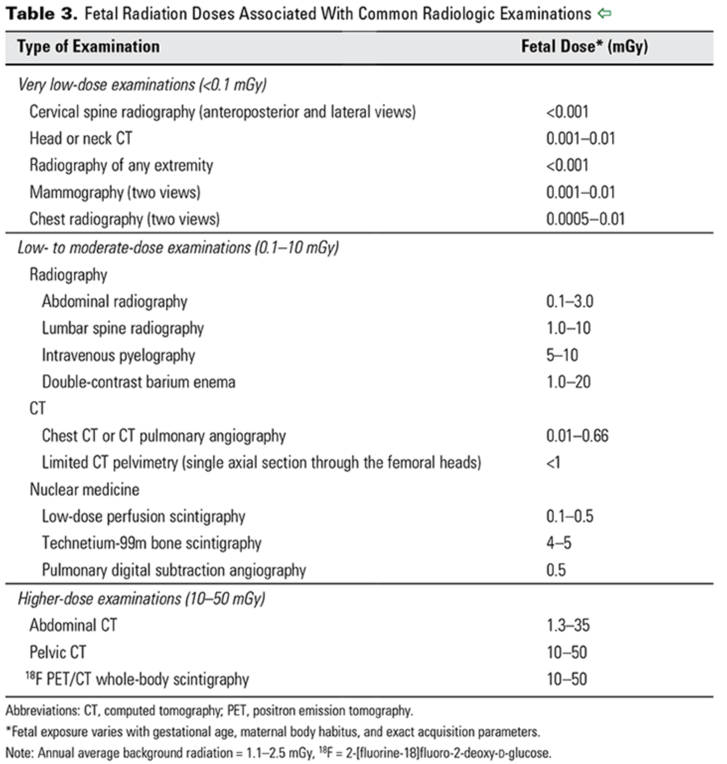
  • The background dose of radiation a fetus is exposed to during pregnancy is around 1 mGy.

  • From CO 723 — a reference for doses associated with different imaging studies.

ACOG CO 723

ACOG CO 723

The risk of radiation exposure on a developing fetus depends on both the dose of radiation, as well as the gestational age at which the exposure occurs.

  • For instance, if an exposure of 50-100 mGy occurs prior to implantation (0-2 weeks post fertilization), there is generally an all or none effect; that is to say, this usually results in miscarriage, or no consequence at all.

  • During organogenesis, or 2-8 weeks post-fertilization, congenital anomalies or growth restriction can be seen with cumulative doses of 200-250 mGy.

  • The risk of severe intellectual deficit or microcephaly is most prominent around 8-15 weeks, with doses between 60 - 300 mGy.

    • There is an estimated 25 point IQ loss per 1000 mGy exposure during this time period. 

    • A lower risk of severe intellectual disability may persist through 25 weeks gestation, though again with exposures of 250mGy or more.

  • Other risks include childhood cancer. With respect to leukemia, it is estimated the risk of childhood leukemia increases 1.5-2 fold with a 10-20mGy dose, over a background leukemia risk of 1 in 3000.

  • Radiologists and radiation physicists can help to calculate doses for patients exposed to multiple studies or with occupational hazards. 

With respect to contrast:

  • Oral contrast poses no real or theoretical harm to pregnant or lactating mothers and their infants.

  • IV contrast tends to be iodinated, but is also water-soluble.

    • So similarly to gadolinium, in pregnant patients this crosses the placenta.

    • Animal studies have demonstrated no teratogenic effects from its use, but it is recommended to limit use of iodinated contrast unless necessary.

    • Also similarly to gadolinium, because of this water solubility, iodinated contrast is excreted minimally in breastmilk, and breastfeeding should be continued without interruption.

Nuclear medicine studies

Radioisotopes for nuclear medicine studies, such as VQ scans, thyroid scans, and bone scans, are variable in their potential effects on the fetus.

  • Technetium-99 is one of the most common radioisotopes used for these studies, and given its short half life of 6 hours as well as its pure gamma ray emission, is generally accepted as safe to use when indicated in pregnancy.

  • Radioactive iodine (I-131), by contrast (punny!), readily crosses the placenta and has a half-life of 8 days, and has known adverse effects on the fetal thyroid. Thus, it is contraindicated for use in pregnancy, and is also recommended against use in breastfeeding mothers until breast milk has been cleared of the radioisotope.