Breast Imaging and Density

Today we welcome Dr. David Edmonson, assistant professor in surgery and obstetrics and gynecology at the Warren Alpert Medical School of Brown University. Dr. Edmonson is an expert in breast disease and a surgical oncologist. Today he talks with us on imaging and breast density.

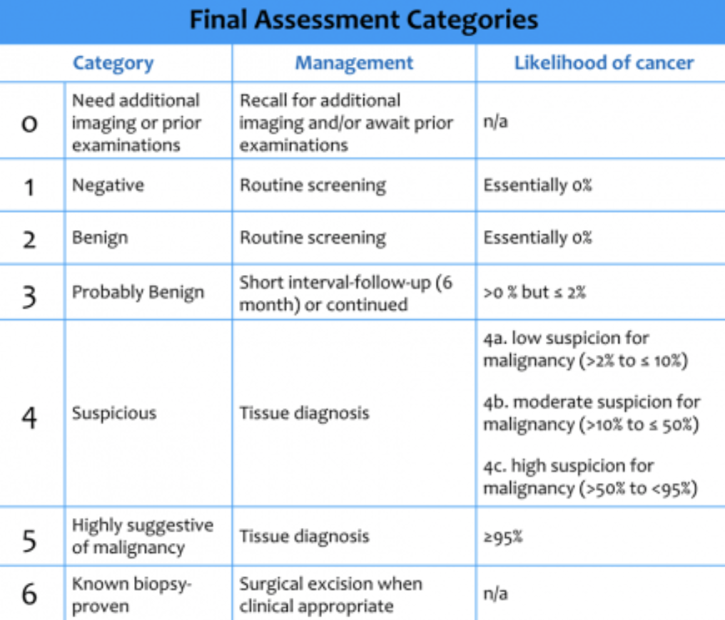
Mammography results can be classified into the BI-RADS (“Breast Imaging Reporting And Data System”) categories — it’s worthwhile to remember these categories and what the likelihood of malignancy is:

(c) Radiology Assistant

(c) Radiology Assistant

Mammography will also often report breast density. Breast density can hinder the utility of mammography, and depending on your area of practice, may suggest or require additional study. Dr. Edmonson does note that breast density requirements do have limited data, but are the subject of active study.

You can use risk models to help assess need for additional imaging: the Tyrer Cuzick or Gail models can be utilized, taking into account different risk factors.

To find out more about breast imaging and density, check out the immensely helpful DenseBreast-Info.org. On their website, there are multiple opportunities to expand your knowledge, including the CME opportunity Breast Density: Why It Matters as well as an FAQ for healthcare providers.

Benign Breast Disease

Today’s episode suggestion comes to us from Shadae Beale, a resident at Meharry Medical College. Thanks for listening! And be sure to send us your ideas through the website or via email!

We kicked off today’s episode with a vignette:

24 yo F G1P1 with 10 year history of type 1 diabetes presents with right breast mass that she palpated in the shower. She is currently breastfeeding her 9 mo old baby. She states that she has had some pain in her right breast, though no redness or swelling, and that she has always had “lumpy breasts.” She is worried because her 78 yo grandmother was recently diagnosed with breast cancer. No other breast or ovarian cancer in her family. On exam, she has lumpy, cobblestone texture of both breasts, with free-moving tissue throughout. There is one 2x2cm firm, mildly tender, mobile mass in the R breast at the 2 o’clock position, approximately 1 cm from the nipple. No axillary lymphadenopathy. 

Now we imagine that if this isn’t a likely scenario in your clinic time, it is on your test prep questions. What do you do with this patient? Let’s first review a broad differential diagnosis for a likely benign mass:

Nonproliferative Breast Lesions 

  • Breast cysts 

    • Simple cysts - benign, fluid filled mass; usually discrete, compressible, or ballotable solitary mass 

    • Galactocele - milk retention cyst common in women who are breastfeeding 

  • Fibrocystic changes - common, especially in premenopausal women; may cause breast pain 

  • Lipoma - mature fat cells 

  • Fat necrosis - can develop after blunt trauma to the breast; can also occur after surgery (ie. breast reconstruction, radiation therapy); associated generally with skin ecchymosis 

  • Breast abscess - localized collection of inflammatory exudate; can develop alongside mastitis or cellulitis; usually will have all the signs of infection! 

  • Diabetic mastopathy 

    • Usually in women with longstanding T1DM 

    • Suspicious fibrous breast lumps, usually multiple 

    • Need to biopsy for diagnosis

  • Idiopathic granulomatous mastitis 

    • Rare inflammatory disease of the breast - usually presents as a painful, firm and ill-defined mass that can have erythema and edema of the skin 

Proliferative Breast Lesions without Atypia 

  • Intraductal papillomas 

    • Monotonous array of papillary cells that grow from the wall of a cyst into its lumen.

    • Most common cause of bloody nipple discharge (key to any vignette!)

    • Generally not concerning, but CAN harbor DCIS; can be solitary or multiple lesions. If bothersome or concern for atypia, surgical excision is performed.

Intraductal papilloma. (C) WebPathology.

  • Sclerosing adenosis - lobular lesion with increased fibrous tissue; no need to treat.

  • Radial scar - complex sclerosing lesions; usually diagnosed after biopsy. Recommend excision, but no other treatment .

  • Fibroadenoma 

    • Most common benign tumor in the breast, accounting for ½ of all breast biopsies 

    • Glandular and fibrous tissue, presenting as a well defined, mobile mass on exam.

Atypical Hyperplasia 

  • Atypical ductal hyperplasia (ADH)

    • Proliferation of uniform epithelial cells with round nuclei fill part of the duct.

    • Standard of care after biopsy-proven diagnosis is surgical excision, due to risk of upgrade to ductal carcinoma.

  • Atypical lobular hyperplasia (ALH)

    • Monomorphic, evenly spaced dyshesive cells fill part of the lobule; can also involve ducts.

    • Referral to breast onc should occur, as management varies based on other clinical risk factors.

OK, so now you have a differential — what do we still need to do for this patient in front of us?

Always starting off with a history is important. With respect the HPI, it’s important to know not only about the characteristic of the mass, but any changes to the mass and the timiing of changes. For instance, is it painful, but cyclically painful with menses? That would argue more for fibrocystic changes. Has it grown in size over the last 3 months and caused nipple inversion in the meantime? That’s more worrisome for malignancy.

Family history and social history are also exceptionally important. Smoking increases risks of certain breast pathologies. And family history is obviously tantamount to determining a patient’s risk for particularly early-onset breast cancer.

Physical examination should include both breasts, examined in both a sitting and recumbent position. Note asymmetry, skin changes, nipple changes, and the location of masses. Generally using clock face language is most helpful for your referral: i.e., “12:00 position, 3cm from nipple” is highly descriptive. Finally, a regional lymph node exam should also be performed. Generally this includes axillary and supraclavicular nodes.

Imaging is what we will turn to next. For the younger patient, targeted breast ultrasound is an excellent choice, as it’s more sensitive than mammogram in this population with denser breast tissue. It also allows for immediate biopsy should the reading radiologist decide it’s indicated. Diagnostic mammography is also a standard of care in anyone with a palpable breast mass who meets criteria for screening. Definitive diagnosis is achieved with biopsy — core biopsy for solid lesions, fine needle aspiration for cysts, or excisional tissue biopsy as another option.