Chronic Pelvic Pain

Today we welcome Dr. Eva Reina, who is a current PGY-3 at Brown/Women and Infants. Eva shares with us some information on one of the most clinically challenging topics in office gynecology: chronic pelvic pain.

In terms of the initial evaluation, using the PPUBS framework can be useful:

  • Pain: typical “can you describe your pain?”

  • Periods: a thorough menstrual history, which should always be part of your GYN history taking.

  • Urinary symptoms: timeline of symptoms with respect to urination, as well as triggers for any symptoms related to urination.

  • Bowels: constipation and diarrhea, depending on pattern, can relay problems such as IBS, IBD, or other functional GI disorders.

  • Sexual function: describing not only the nature of dyspareunia, but the timing and length of pain symptoms.

After history taking, a physical examination is the next important step. Particularly with the pelvic exam, starting with a single digit may be revelatory. Deep infiltrating endometriosis may be suggested by particular point tenderness. Bimanual examination can help to assess uterine size or concerns for adenomyosis. Speculum exam may not reveal much in the way of pain, but allows for testing for infectious etiologies.

We then discuss some of the mechanisms of chronic pelvic pain development:

Central Sensitization: Most patients with chronic pelvic pain do have multiple pain generators and may even be centrally sensitized: that is, amping up of a stimulus leading to a state of constant reactivity. This lowers the threshold of a stimulus to cause pain and also may maintain pain even after the initial insult has been removed. For instance, in population-based studies vulvodynia, IC, endo, IBS, migraines are commonly found together.

Cross-Talk:

  • The top-down phenomenon. The patient who has low back pain, migraines, fibromyalgia, and has now been referred to your office with chronic pelvic pain. 

  • The bottom-up phenomenon. 30 year old female who has “always had painful periods.” Stopped OCPs 5 years ago in order to achieve pregnancy. Presenting to your office for urinary symptoms (pain with bladder filling, urgency, frequency). 

  • Pelvic Organ Cross-Sensitization: Convergence of sensory input from the pelvic viscera, their associated striated sphincters, muscular components of the pelvic floor, lower abdominal wall, the muscular perineum and its corresponding cutaneous components. 

Afferent information from the major pelvic organs (e.g. bladder, bowels, and uterus) is conducted through the hypogastric, splanchnic, pelvic, and pudendal nerves to cell bodies in thoracolumbar and lumbosacral dorsal root ganglia. At first, these signals go up to the brain (central nervous system), and an efferent signal comes back down (e.g. reflexically removing your hand from a hot stove). 

Over time, if one doesn’t remove the noxious stimulus, the body thinks there must be nobody home to hear the fire alarm, let’s try the neighbor’s house. So, long-term, noxious afferent stimulation from an irritated pelvic organ (peripheral to central) leads to that sensory input traveling back down to the peripheral nervous system to pass the message along to a nearby pelvic organ that was not previously affected. This neurogenic “inflammation” via the central to peripheral pathway may even produce functional changes in the uninsulted organ with little evidence of an organic etiology (e.g. IBS, IC/PBS, vulvodynia). 

Where do the muscles come in? 

  • It is helpful to think of muscular spasm as a reactive phenomenon. If you uncover pelvic or abdominal wall myalgia, be sure to treat other primary pain generators BEFORE pelvic floor PT. If you don’t remove the stimulus, the patient is unlikely to make sustained forward progress in PT. 

  • Pelvic floor PT is hard to come by (shortage of well-trained providers) and it is uncomfortable both physically and mentally for patients. Thus it is difficult to motivate patients to return to pelvic floor PT after a previous failure of therapy. 

Breast Cancers and Treatment Knowledge for the OB/GYN

Today we welcome back Dr. Edmonson for part II of our breast cancer chat. Check out last week’s post for screening and imaging information.

From a radiographically-guided core breast biopsy, there’s a lot of things that can come back. Today we’ll focus on the pathologic concerns. Dr. Edmonson breaks this down into:

Atypical Ductal Hyperplasias / Atypical Lobular Hyperplasias / Lobular Carcinoma in Situ
These lesions predict a risk for breast cancer in the future, but are not actually cancer. These are all managed surgically, with an approximately 15% upstaging rate on final pathology after excision, most pronounced with ADH. The risk models we discussed last episode can then give an updated risk of breast cancer which may alter screening strategy (i.e., if risk exceeds 20%, MRI may be used as an adjunct). Additionally, using risk-reducing medications such as tamoxifen or raloxifene may also become appropriate.

Ductal Carcinoma in Situ (DCIS)
These are non-invasive cancers which require lumpectomy or potentially mastectomy. Thereafter, radiation is usually recommended as well as risk-reducing medications such as tamoxifen or aromatase inhibitors such as anastrozole. There is a 20-30% upstaging risk at time of surgery, and additionally additional surgery may be required to get negative surgical margins as there is no reliable intraoperative technique to detect margins.

Invasive Cancers (Invasive Ductal Carcinoma or Invasive Lobular Carcinoma)
With invasive cancer, different strategies exist. Surgery versus neoadjuvant chemotherapy or endocrine therapy may be considered based on extent of disease to reduce morbidity of surgery (i.e., more limited lymph node dissection which would reduce risk of lymphedema from axillary dissection). New surgical techniques exist now as well including lymphatic reanastamosis that is helping to improve morbidity after these surgeries.

Invasive lobular carcinoma can be difficult to identify. These are less aggressive, but they often don’t show up well on imaging and are difficult pre-operatively to get good margins.

After A Diagnosis
OB/GYNs can help to reassure patients and connect them to breast surgeons. Fewer breast cancers are requiring chemotherapy, which is often a patient’s greatest fear. Surgical techniques are improving and reconstruction is widely available, including skin-sparing and nipple-sparing techniques.

Screening will be guided by the breast surgeon. This depends more and more on the individual, the pathology and tumor characteristics, and risk for local recurrence. Recall that patients on tamoxifen are at higher risk of VTE and at higher risk of endometrial hyperplasia.

Breast Imaging and Density

Today we welcome Dr. David Edmonson, assistant professor in surgery and obstetrics and gynecology at the Warren Alpert Medical School of Brown University. Dr. Edmonson is an expert in breast disease and a surgical oncologist. Today he talks with us on imaging and breast density.

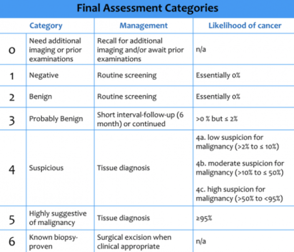
Mammography results can be classified into the BI-RADS (“Breast Imaging Reporting And Data System”) categories — it’s worthwhile to remember these categories and what the likelihood of malignancy is:

(c) Radiology Assistant

(c) Radiology Assistant

Mammography will also often report breast density. Breast density can hinder the utility of mammography, and depending on your area of practice, may suggest or require additional study. Dr. Edmonson does note that breast density requirements do have limited data, but are the subject of active study.

You can use risk models to help assess need for additional imaging: the Tyrer Cuzick or Gail models can be utilized, taking into account different risk factors.

To find out more about breast imaging and density, check out the immensely helpful DenseBreast-Info.org. On their website, there are multiple opportunities to expand your knowledge, including the CME opportunity Breast Density: Why It Matters as well as an FAQ for healthcare providers.

Group B Strep

Today’s episode covers the ACOG Committee Opinion on Group B Streptococcus, also known as GBS or Streptococcus agalactiae.

The OBG Project has a phenomenal summary of these new guidelines; we encourage you to check out their outline which was hugely helpful in preparing the podcast for today! And be sure to check out how you can get their premium product, OBG First, absolutely FREE if you’re a chief resident. Check out the sidebar to learn more!

Get to know Group B Strep

Streptococcus agalactiae or GBS is a common flora of the vagina and rectum of women, with a prevalence of colonization between 10-30%.  In newborns, two types of GBS-related disease exist:

  • Late Onset Disease occurs between 7 days and 2-3 months after birth, and is characterized by sepsis, more commonly meningitis, and organ/soft tissue infection.

  • Early Onset Disease is present within 7 days of birth.

    • Occurs secondary to vertical transmission; fetal/neonatal aspiration during labor process; or both. 

    • Manifestations of disease occur most likely within 12-48 hours after birth. 

    • Characterized by sepsis, pneumonia, and less commonly meningitis.

    • The most common cause of early-onset neonatal sepsis.

      • 72% of cases of Early Onset Disease occur in term newborns; however, mortality is markedly higher in premature infants (19.2% in premies vs 2.1% at term), and premature infants are more likely to require higher levels of intensive care intervention. 

    • When treating GBS in labor, this is what we are primarily aiming to prevent! 

    • Risk factors for GBS Early Onset Disease:

      • Gestational age < 37 weeks

      • Very low birth weight

      • Prolonged rupture of membranes > 18 hours

      • Intraamniotic infection

      • Young maternal age

      • Black race

      • Heavy vaginal-rectal colonization or GBS bacteriuria (proxy for heavy colonization)

      • Previous newborn affected by GBS early onset disease

The Screening Recommendations

  • Screening in the USA is universal, meaning this should be a standard part of prenatal care. 

    • ACOG recommends universal screening for vaginal-rectal colonization between 36’0 - 37’6.

      • These results are considered “valid” for 5 weeks — thus the majority of women will be captured with screening at 36’0 (assuming inductions for most women at 41’0). 

      • This is also recommended for women planning to deliver by cesarean.

  • What about GBS bacteriuria?

    • If GBS bacteriuria is present in any amount at any time during pregnancy, this is considered a positive result (proxy for heavy colonization), and thus repeat screening is not necessary.

    • If asymptomatic GBS bacteriuria is present at >10^5 CFU/mL, treatment should be considered as you would for any other form of ASB. 

      • If at less than 10^5 CFU, no correlation has been found between treatment of this lower-level bacteriuria and improved maternal or neonatal outcomes; however, it should still be noted that this patient would be considered GBS positive.

  • Screening may also occur at the time of admission for preterm labor and prelabor premature rupture of membranes (PPROM)

    • In the case of prematurity, treatment should begin while awaiting screen results.

      • If the screen is positive and preterm labor resolves, the colonization should be considered positive for the remainder of the pregnancy.

      • If the screen is negative, re-screening should be performed after 5 weeks. 

  • The gold standard for screening is obtaining a vaginal-rectal culture. 

    • Recently, nucleic acid amplification tests (NAAT) have also demonstrated promise in obtaining accurate, quick results.

      • Most accurate tests that perform similar to culture do require some culture time prior to the NAAT test performance; results generally aren’t available before 18-24 hours after screening occurs. 

        • NAAT tests that offer results in a shorter time period often have lower accuracy.

      • NAAT tests also do not have the ability to test antibiotic resistance, which is important in the case of a patient with known penicillin allergy.

        • If your patient has known penicillin allergy, the laboratory should be alerted so sensitivity testing can be performed.

    • Given this delay and with someone moving quickly in labor or preterm labor with no known GBS status, risk factor-based screening may also be employed to decide if treatment should be pursued in the absence of a screening result. 

      • If any of the risk factors are present, treatment should be pursued:

        • Prematurity or PPROM (< 36 weeks 6 days)

        • History of a prior newborn affected by GBS disease

        • Amniotic membrane rupture > 18 hours duration

        • Presence of intrapartum fever > 100.4F (38 C)

        • If known GBS positive result in a previous pregnancy (may engage in shared decision making in this clinical scenario).

Intrapartum Antibiotic Treatment 

  • The gold standard for the treatment of GBS colonization intrapartum to reduce risk to neonates for early-onset disease is penicillin G. 

    • Dosed at a loading dose of 5 million units loading, then 2.5-3 million units IV every 4 hours until delivery.

    • Ampicillin is an acceptable alternative, with a loading dose of 2g IV followed by 1g IV every 4 hours. 

      • PCN is preferred as it has a narrower, more targeted spectrum of activity and lower likelihood of inducing resistance in other organisms.

    • For this reason, identification and history of penicillin allergy is super important to flush out in prenatal care! 

      • 80-90% of persons with reported PCN allergy are not truly allergic. 

        • Pruritic rash, urticaria (hives), immediate flushing, angioedema, respiratory distress, or anaphylaxis after PCN or cephalosporin administration is considered a high risk for true allergy.

        • Even with high risk persons, if PCN allergy testing has not been performed, it is recommended to do this — even in pregnancy!

    • When PCN allergy testing has not been performed, the allergy should be classified as “low” versus “high risk,” based on the symptoms just described.

      • If “low risk,” first generation cephalosporins such as cefazolin are recommended. 

        • Cefazolin has very low cross reactivity with penicillin for a 1st gen cephalosporin, and GBS remains highly susceptible to it. 

        • Dosed at 2g IV load, followed by 1g IV q8h until delivery.

      • If “high risk,” or an unknown risk with no allergy testing, susceptibility testing should be performed by the laboratory with screening.

        • Options for therapy will include clindamycin or vancomycin.

          • Clindamycin should only be utilized if culture results have shown susceptibilit.

            • Resistance to clindamycin in GBS is approximately 20%. 

            • Dosing: 900mg q8h until delivery 

          • Vancomycin has good activity against GBS.

            • High risk in creating resistant organisms (i.e., VRE) with widespread use, and thus every effort should be made to rule out penicillin allergy before its use.

            • Dosing: 20mg/kg IV q8h, with maximum of 2g per single dose.

  • How long does it take the antibiotics to work?

    • All antibiotics used for GBS prophylaxis are time-dependent with respect to their ability to lower microbial load.

      • Studies done with PCN or ampicillin prophylaxis demonstrate that 4 or more hours pf prophylaxis is preferable, though 2 hours has been shown to reduce GBS count and decrease neonatal sepsis. 

      • That said, obstetric intervention should not be delayed solely to provide 4 hours of antibiotic administration., when it is indicated.

        • Examples of such interventions that should not otherwise be delayed include oxytocin administration, AROM, or cesarean.

That said, if interventions are not immediately indicated, the benefits of increased antibiotic exposure to reduce GBS exposure should be considered.

Bacteruria, UTI, and Pyelonephritis

Today, we’re going to review all the iterations of urine culture abnormalities in pregnancy. We screen urine cultures in the first trimester and many times again later on in pregnancy, and urinary symptoms are a common complaint.

Asymptomatic bacteriuria is when high levels of bacteria are in urine without associated symptoms. This occurs in 2-7% of pregnant women and typically occurs early in pregnancy.

Urinary tract infections can be broken down into two sub-categories:

  1. Lower UTI, or acute cystitis is basically an infection of the bladder, with symptoms of dysuria, urinary frequency, and urgency. They occur in 1-2% of pregnant women.

  2. Upper UTI, or acute pyelonephritis may have symptoms of simple cystitis, but include systemic symptoms of infection: flank pain, fevers, chills, nausea/vomiting and costovertebral angle (CVA) tenderness. They occur in 0.5-2% of pregnant women, and more commonly in the 2nd and 3rd trimesters.

Non pregnant women can experience asymptomatic bacteriuria as well as pregnant women. In the non pregnant patient, ASB can be quite prevalent, particularly as we age — over 20% in patients >80 years old. Multiple studies have shown that treatment of asymptomatic bacteriuria in the general, nonpregnant population does not reduce frequency of symptomatic infection or prevent adverse outcomes. Importantly, adhering to this principle helps with antibiotic stewardship.

Pregnant women, however, are different story. As many as 20-35% of pregnant women with asymptomatic bacteriuria will develop symptomatic cystitis or pyelonephritis if untreated, and risk is reduced by 70-80% with treatment. Asymptomatic bacteriuria is not only associated with pyelo, but also associated with adverse pregnancy outcomes, like preterm birth and low birth weight infants. Thus, versus other populations, ASB treatment is imperative in pregnancy.

The most common organisms implicated in these urinary infections include: 

  • E. coli - about 70% 

  • Klebsiella  - about 3% 

  • Enterobacter - about 2% 

  • Gram positive organisms (like GBS) = 10% 

Diagnosis and Screening Recommendations for ASB, Cystitis, and Pyelonephritis:

  • Asymptomatic bacteriuria 

    1. Finding high-level bacterial growth on urine culture without symptoms consistent with a UTI.

    2. Recommendation per Infectious Disease Society of America to screen all pregnant women for asymptomatic bacteriuria at least once in early pregnancy (usually 12-16 weeks).

    3. Diagnostic criteria:

      1. Isolation of same bacterial strain greater than or equal to 10^5 colony-forming units/mL in two consecutive voided urine samples, or

      2. A single catheterized specimen with one bacterial species isolated in >/= 10^2 cfu/mL 

  • Acute Cystitis 

    1. Symptoms as described above (dysuria, etc) with pyuria seen on urinalysis.

    2. Should be confirmed with urine culture, but with typical symptoms and pyuria, should start treating without the culture coming back.

    3. Cystitis should NOT involve systemic symptoms.

  • Pyelonephritis 

    1. Can have all of the above (though dysuria may not always be present).

    2. Fevers, chills, flank pain, nausea/vomiting, CVA tenderness.

    3. Pregnant women can become VERY sick 

      1. Estimated that as many as 20% of pregnant women with pyelonephritis develop complications like septic shock syndrome or variants like ARDS.

      2. One study of 32,282 pregnant women in the general obstetric population with pyelo → 23% had anemia, 17% had bacteremia, 7% had respiratory insufficiency, and 2% had renal dysfunction.

    4. Diagnostic evaluation can include:

      1. Urinalysis, urine culture.

      2. CBC

      3. Possibly blood cultures and lactic acid if patient presents with sepsis.

      4. Imaging not routine, but in patients who are severely ill or who have symptoms of renal colic, diabetes, prior urologic surgery, immunosuppression, urosepsis, or repeated pyelo, imaging can help identify other complicating factors, ie. infected stone, renal abscess.

      5. Can consider CT, but ultrasound preferred due to decreased radiation exposure in pregnant women.

Treatment 

  • Asymptomatic bacteriuria 

    1. Basic principle is to treat with antibiotics tailored to culture results and then follow up cultures to confirm sterilization of the urine.

    2. Possible antibiotics to consider include beta-lactams, nitrofurantoin, or fosfomycin.

    1. A short course is usually effective in eradicating asymptomatic bacteriuria,  although a single-dose regimens may not be as effective as slightly longer regimens. However, optimal duration of therapy is uncertain.

      1. The only exception is fosfomycin - a single dose can successfully treat bacteriuria.

    1. Follow up 

      1. Up to 30% of women fail to clear asymptomatic bacteriuria after short course therapy, so repeat culture is recommended about a week after finishing antibiotics.

      2. However, there isn’t a lot of data about if we should repeat another culture later on for repeat screening after treatment, or if this is even necessary.

      3. Similarly, not a lot of data about treating again if repeat culture is positive and for how long, though general consensus is to treat.

  • Acute cystitis 

    1. Treatment is usually empiric because it’s hard to make someone wait until cultures are back.

    2. Same antibiotic treatment options as for asymptomatic bacteriuria.

    3. Again, treatment time is uncertain, but usually 3-7 day course as long as there are no symptoms/signs of pyelo (except fosfomycin, which is single-dose regimen).

    4. Follow-up repeat culture 7 days after antibiotic completion, as with ASB.

    5. If a woman has 2 or more episodes of recurrent cystitis in pregnancy, it is reasonable to start antibiotic prophylaxis:

      1. Cephalexin 250-500mg qHS or nitrofurantoin 50-100mg qHS depending on susceptibility of organisms on previous cultures.

  • Pyelonephritis 

    1. Pregnant people need to be admitted because they are WAY more likely to get super sick compared to nonpregnant women.

    2. Parenteral antibiotics initially, then converted to oral antibiotics when woman has been afebrile for 24-48 hours.

    3. Empiric antibiotics:

      1. Broad spectrum beta-lactams - ie. ceftriaxone 

      2. If someone has ESBL (extended spectrum beta-lactamase) bacteria, can consider a carbapenem.

      3. Avoid fluoroquinolones and aminoglycosides in pregnancy.

    4. Oral antibiotics:

      1. Should usually be beta-lactams, or if second trimester, can consider trimethoprim-sulfamethoxazole (Bactrim).

      2. Do not use nitrofurantoin or fosfomycin because they do not achieve adequate levels in the kidney.

    5. Suppression/Prophylaxis:

      1. Should consider preventative therapy for the duration of pregnancy because recurrent pyelo during pregnancy occurs in 6-8% of women.

      2. Some practices continue antibiotic prophylaxis for six weeks postpartum, but the data is unclear to the benefit of this.