Diagnostic Imaging During Pregnancy and Lactation

Today we’re going to review a source of constant consultation and confusion: diagnostic imaging during pregnancy and breastfeeding. ACOG CO 723 is the definitive reading on this subject, and we use it to structure this episode. Critical take home: ACOG states that critical imaging studies should not be withheld from a pregnant patient if needed to make a diagnosis. 

Ultrasound

  • Sonography utilizes sound waves to produce a visible image, and is not a form of ionizing radiation.

    • Thus, it is considered the safest mode of imaging in pregnancy.

    • However, ACOG still recommends sticking to the ALARA principle of exposure in pregnancy (“As Low As Reasonably Achievable”) to minimize any potential untoward effects. 

    • One of these theoretical effects involves color or spectral flow Doppler. Due to its intensity, the theoretical temperature increase surrounding the area being study can be as high as 2deg C, or 3.6deg F. It’s unlikely than any temperature increase would be sustained at any fetal anatomic site to cause harm. However, for this reason, even ultrasound exposure should be used judiciously. 

MRI

  • Allows for visualization of soft-tissue structures like ultrasound

    • However, as MRI is operator-independent, pick up rates for certain pathologies like appendicitis tend to be higher.

    • There are no special contraindications or considerations in pregnancy for non-contrast MRI, other than the usual screening surrounding metal or magnet-sensitive implants, such as pacemakers. 

  • Non-contrast MRI is sufficient for diagnosis; however, some diagnoses or studies may be improved by the use of gadolinium-based contrast, for which there is uncertainty regarding fetal effects.

    • Gadolinium is water-soluble, and thus crosses the placenta into fetal circulation.

    • Free gadolinium is toxic, so it is bound, or chelated, when administered for studies.

      • There is concern that since this bound gadolinium can enter fetal circulation, it can recycle in the fetal circulation. This potentially could sit for long enough that the gadolinium could dissociate and become free; thus become toxic. 

      • Given at least the concern for potential poor outcomes, gadolinium-based contrast should be limited in use to cases where there is an absolute clear benefit to its administration.

    • Gadolinium’s water-solubility makes it an OK contrast agent to use during lactation.

      • Less than 0.04% of a dose of gadolinium will be excreted in breastmilk in the first 24 hours, and less than 1% of this will be absorbed in the infant GI tract. Thus, breastfeeding should not be interrupted after gadolinium contrast studies.

CT, XRAY, and other ionizing radiation studies
Before talking about ionizing radiation studies, it’s important to know some vocabulary and measurements of radiation:

  • Exposure is the number of ions produced by radiation in the form of X-rays or gamma rays per kilogram of air. This is measured in Roentgen units.

  • Dose is the amount of energy deposited per kilogram of tissue. This is the usual consideration when we talk about radiation in pregnancy. This is measured in rads or in Gray units; 100 rad is equivalent to 1 Gray.

  • Relative effective dose is the amount of energy deposited per kilogram of tissue, and normalized for biological effectiveness on the tissue. This is measured in Roentgen equivalent men (rem) or Sievert units.


Again, the dose is what we usually consider and track with respect to radiation in pregnancy.

  • The background dose of radiation a fetus is exposed to during pregnancy is around 1 mGy.

  • From CO 723 — a reference for doses associated with different imaging studies.
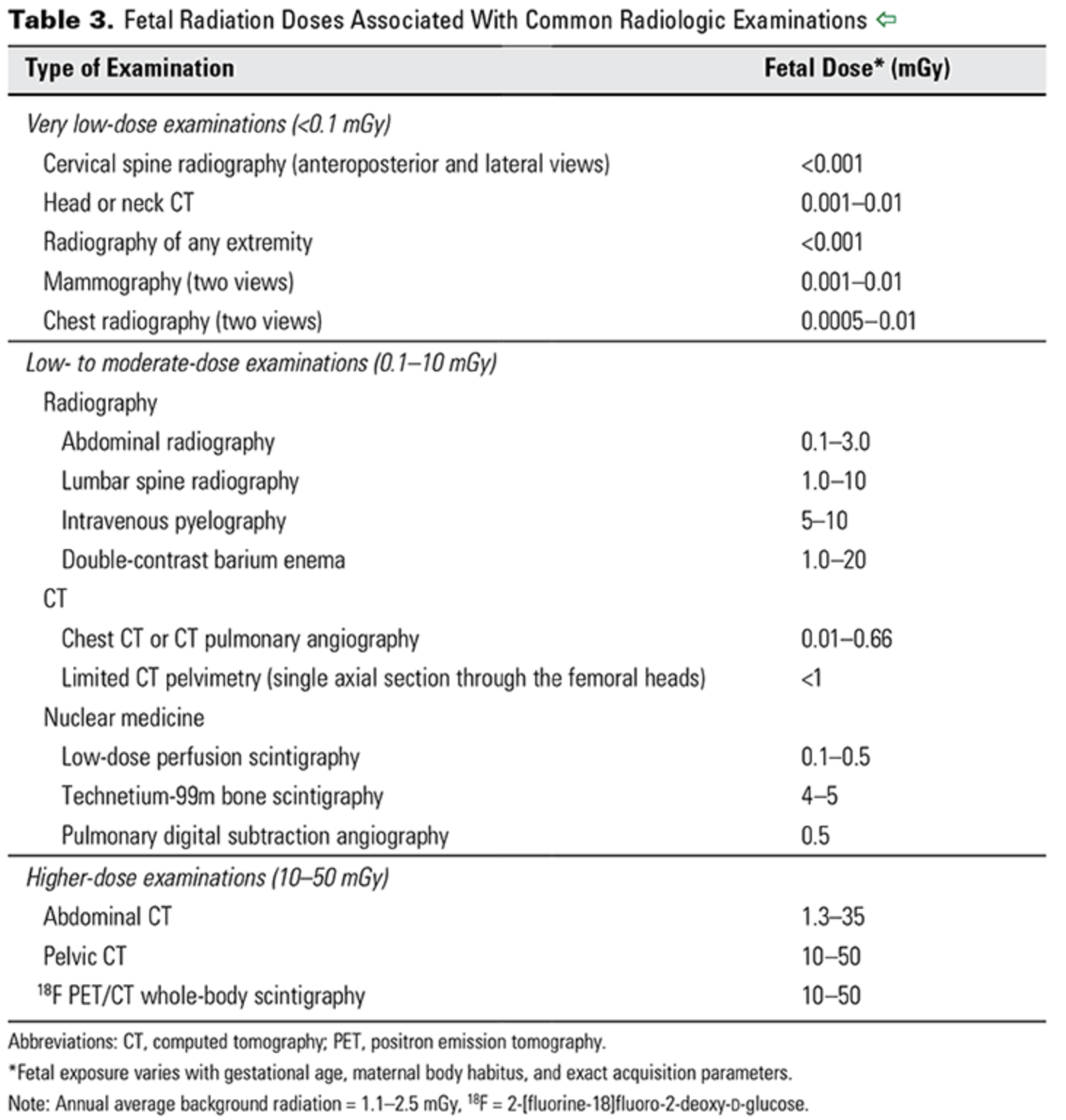
ACOG CO 723

ACOG CO 723

The risk of radiation exposure on a developing fetus depends on both the dose of radiation, as well as the gestational age at which the exposure occurs.

  • For instance, if an exposure of 50-100 mGy occurs prior to implantation (0-2 weeks post fertilization), there is generally an all or none effect; that is to say, this usually results in miscarriage, or no consequence at all.

  • During organogenesis, or 2-8 weeks post-fertilization, congenital anomalies or growth restriction can be seen with cumulative doses of 200-250 mGy.

  • The risk of severe intellectual deficit or microcephaly is most prominent around 8-15 weeks, with doses between 60 - 300 mGy.

    • There is an estimated 25 point IQ loss per 1000 mGy exposure during this time period. 

    • A lower risk of severe intellectual disability may persist through 25 weeks gestation, though again with exposures of 250mGy or more.

  • Other risks include childhood cancer. With respect to leukemia, it is estimated the risk of childhood leukemia increases 1.5-2 fold with a 10-20mGy dose, over a background leukemia risk of 1 in 3000.

  • Radiologists and radiation physicists can help to calculate doses for patients exposed to multiple studies or with occupational hazards. 

With respect to contrast:

  • Oral contrast poses no real or theoretical harm to pregnant or lactating mothers and their infants.

  • IV contrast tends to be iodinated, but is also water-soluble.

    • So similarly to gadolinium, in pregnant patients this crosses the placenta.

    • Animal studies have demonstrated no teratogenic effects from its use, but it is recommended to limit use of iodinated contrast unless necessary.

    • Also similarly to gadolinium, because of this water solubility, iodinated contrast is excreted minimally in breastmilk, and breastfeeding should be continued without interruption.

Nuclear medicine studies

Radioisotopes for nuclear medicine studies, such as VQ scans, thyroid scans, and bone scans, are variable in their potential effects on the fetus.

  • Technetium-99 is one of the most common radioisotopes used for these studies, and given its short half life of 6 hours as well as its pure gamma ray emission, is generally accepted as safe to use when indicated in pregnancy.

  • Radioactive iodine (I-131), by contrast (punny!), readily crosses the placenta and has a half-life of 8 days, and has known adverse effects on the fetal thyroid. Thus, it is contraindicated for use in pregnancy, and is also recommended against use in breastfeeding mothers until breast milk has been cleared of the radioisotope. 


Management of an Early Unlocated Pregnancy

Today we’re bringing back Dr. Erin Cleary one more time before she transitions to her new role as an MFM fellow at the Ohio State University! Dr. Cleary today talks with us on early pregnancy of unknown location - a common problem in the office or the emergency department/triage.

Women presenting to the ED with first trimester bleeding, pain, or both, have had a demonstrated prevalence rate of ectopic pregnancy up to 18% in some studies. Ruptured ectopic is a leading cause of pregnancy-related mortality in the first trimester, accounting for 2.7% of pregnancy-related deaths overall in 2011-2013. Proper identification and management of early, unlocated pregnancy is life-saving!

Dr. Cleary was kind enough to put together her high points from this episode for our blog post today:

H&P:

  • Any patient with an unlocated pregnancy should be considered to have a potential ectopic pregnancy.

    • Women with prior ectopic, regardless of method of treatment, are at risk for ectopic in a subsequent pregnancy (three- to eightfold higher compared with other pregnant women).

    • If pregnancy is present while IUD is in place, risk of ectopic is 1 in 2 pregnancies for the levonorgestrel IUD and 1 in 16 pregnancies for the copper IUD.

    • Women with a history of PID have an approximately threefold increased risk of ectopic pregnancy

  • Pelvic exam. THIS MUST BE DONE.

Beta-HCG

  • The threshold for a positive qualitative β-hcg test is 20-50 milli-int units, depending on test. For quantitative serum tests, the threshold is 5-10 milli-int units, and 1-2 milli-int units, for ultrasensitive tests.

  • The β-hcg concentration doubles every 29 to 53 hours during the first 30 days after implantation of a viable, intrauterine pregnancy.

  • When ectopic pregnancy is on the differential, a qualitative test is not sufficient. A serum quantitative value is essential to:

    • 1. Interpret imaging (“discriminatory zone”)

    • 2. Have a baseline in the event the β-hcg must be trended

The Discriminatory Zone

  • Definition: A concept that there is a quantitative β-hcg level above which the landmarks of a normal intrauterine pregnancy (yolk sac and embryo) should be visible on ultrasound.

    • Therefore, the absence of a gestational sac when β-hcg level is above the DZ is strongly suggestive of nonviable pregnancy, with 50-70% being ectopic.

  • Pelvic ultrasound is the gold standard first line imaging modality in early pregnancy and for evaluation of suspected ectopic pregnancy

  • Imaging results will fall into 1 of 5 main categories

    • IUP with normal adnexa. Normal pregnancy!

    • IUP with abnormal adnexa. Although rare, must evaluate for heterotopic pregnancy, or presence of both an intra and extra-uterine pregnancy.

    • No IUP, extra-uterine mass with YS/FP. Confirms ectopic pregnancy.

    • No IUP, adnexal mass without YS/FP. Suspicious for ectopic pregnancy

    • No IUP, normal adnexa. Differential includes normal but early IUP, failed IUP, or unidentified ectopic.

  • A patient with a confirmed ectopic requires evaluation and counseling by an OBGYN to evaluate candidacy for medical or surgical evaluation.

Management:

  • Expectant management: serial quantitative β-hcg level assessment ~q 48 hours, only for stable patients.

    • Scenario A: The β-hcg level rises appropriately (doubles approximately every 2 days).

    • Scenario B: The β-hcg level falls precipitously.

    • Scenario C: The β-hcg level neither rises appropriately nor drops precipitously. Now we should be MORE concerned about ectopic pregnancy, but abnormal IUP is also on the differential.

  • Repeat pelvic imaging is very helpful

  • Every patient who is stable and an appropriate candidate to trend β-hcg levels will eventually declare herself, with either a located IUP, a failed IUP/SAB, or a confirmed or presumed ectopic pregnancy.

We will cover ectopics for surgical and medical management in a future episode, so stay tuned!

Emergency Contraception

Today we spend some time with Dr. Leanne Free, who is one of Fei and Nick’s co-residents. As a rising PGY-4 at Brown, Leanne is interested in family planning fellowship and shares some of that interest with us today by talking emergency contraception!

Leanne breaks down for us the main types of emergency contraceptives — the copper IUD and pills. Much of the information Leanne shares is really nicely prepared in graphical format on the BedSider website:

One crazy thing we learned: many levonorgestrel EC formulations are available on Amazon! Though buyer beware, as there have been some news stories regarding these to be potentially expired medicines. Additionally, as Leanne states, EC is most effective immediately after unprotected intercourse, rather than the 48 hours it takes for Prime delivery. All levonorgestrel EC should be available over-the-counter without restrictions for purchase based on age, gender, or parental consent.

Additionally, patients can follow the Yuzpe method by taking birth control pills that they may already have at home. This can be useful for patients who for some reason do not have access to the emergency contraceptives we refer to in the podcast — though an annual visit is a great time to review and prescribe these options!

Espresso: Treatment of Acute Hypertension in Pregnancy and Postpartum

Our second espresso episode focuses on the acute treatment of severe-range BPs in the pregnant and postpartum patient. More or less, we let the freshly released ACOG CO 767 speak for itself.

Below you’ll find the algorithms we describe in the podcast, which are present in ACOG CO 767. In addition to the below, always remember:

-Obtain IV access and labs (CBC, Creatinine, AST, ALT, urine protein:creatinine ratio) for any newly diagnosed patient with severe-range pressures.
-Avoid labetalol in patients with known asthma, as the beta-blockade effect can trigger respiratory issues, as well as those with CHF or pre-existing cardiac disease. Labetalol may also cause neonatal bradycardia due to beta-blockade.
-Immediate-release nifedipine should not be administered sublingually due to possibility of developing precipitous hypotension. Similarly, parenteral hydralazine may also cause precipitous maternal hypotension.
-IV magnesium sulfate should be given at a 4g or 6g bolus initially, followed by 2g/hr drip for the prevention of eclamptic seizures, if not previously given. Adjusted dosing may be required if renal insufficiency is noted on laboratories. Magnesium sulfate is not an antihypertensive agent.

Espresso: Medical Management of Postpartum Hemorrhage

Welcome to our first Espresso Episode! Just like an espresso, this should be a short, sweet, but highly caffeinated review of more familiar topics. These are intended for rapid-fire review — perfect for while you’re running up to that postpartum hemorrhage!

In today’s episode, we really just stick to the medication management for postpartum hemorrhage, though as anyone with experience with these might remember, there are a lot more components than just these medicines to make hemorrhage management successful. That said, an exam, bimanual massage, and uterotonic agents will resolve many of the cases you’ll see on the floor. More important for CREOGs are likely the dosing and side effects of these medicines, which we also review today. The ACOG PB 183 table on these medicines is also below for visual learners.

For when you have a bit more time to sit and breathe after the run up the stairs, check out ACOG PB 183 to review postpartum hemorrhage in full (ACOG membership required).

ACOG PB 183