2nd and 3rd Trimester Bleeding
/Placenta previa - when the placenta partially or totally covers the internal cervical os. Defined as edge of placenta <10 mm from internal cervical os
Occurs approximately 4/1000 births, but varies world wide. Increased risk associated with history of previous placenta previa, previous C-section, and multiple gestation
Approximately 90% of placenta previa identified on ultrasound <20 weeks → resolve before delivery
Painless vaginal bleeding can occur up to 90% of persistent cases
10-20% of women present with uterine contractions, pain, and bleeding
Why we care: can lead to catastrophic bleeding, need for transfusion, and delivery. Can lead to stillbirth
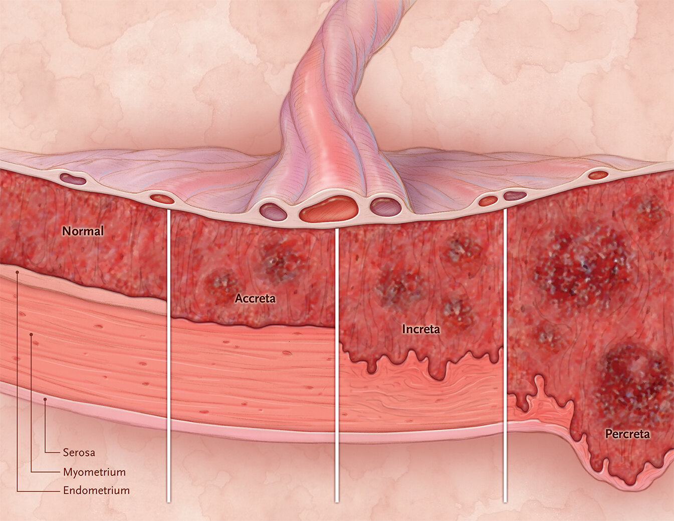
Placenta accreta spectrum
Listen back to our PAS series from March with Drs. Shainker and Einerson:
What it is: abnormal trophoblastic invasion of part of or all of the placenta into the myometrium of the uterine wall
Can be painful or painless bleeding
Risk factors: previous cesarean section, placenta previa, unexplained elevation in MSAFP (but not a good predictor!)
Why we care: can lead to catastrophic bleeding, need for transfusion, and delivery. Also may require hysterectomy. Usually may need delivery at tertiary care center
Vasa previa
What it is: when fetal vessels run within the membranes over the internal os of the cervix
Very rare. Has been quoted 1/2500 deliveries
Painless bleeding usually
Two types:
Velamentous cord insertion and fetal vessels that run freely within the amniotic membranes overlying the cervix or in close proximity of it (2 cm from os); usually pregnancies with low lying placenta or resolved placenta previas are at risk
Succenturiate lobe or multilobe placenta and fetal vessels connectin both lobes course over or in close proximity of cervix (2 cm from os)
Other risks: IVF
Why we care: increased risk of fetal hemorrhage, exsanguination, and death
Placental abruption
What it is: Separation of the placenta from the inner wall of the uterus before birth
Usually painful bleeding
Incidence: 2-10/1000 births in the US
Risk factors: hx of fabruption, cocaine use, tobacco use, hypertension, uterine abnormalities (ie. fibroids, bicornuate uterus)
Why we care: can lead to catastrophic bleeding, need for transfusion, and delivery. Can lead to stillbirth.
Uterine rupture
What it is: significant uterine disruption. Usually will occur along a previous uterine scar
Very painful bleeding (pain is usually more significant than bleeding)
Risk factors: previous uterine rupture, previous uterine scar, especially if a fundal or vertical scar (ie. cesarean delivery, myomectomy), induction, labor
Why we care: very high incidence of morbidity and mortality for both mom and baby
Less dangerous causes:
Labor - “bloody show” with labor
Cervicitis
Can be caused by infection (ie. BV, candida infection, trichomonas, chlamydia, gonorrhea)
Cervical polyp
Vaginal laceration
Doing a Workup for Bleeding in the 2nd and 3rd Trimester
History
How much bleeding? (soaking through clothes? Passing clots?)
Passing tissue?
Remember: just because someone has light bleeding does not mean that they don’t have something life-threatening for them or their fetus
Is there pain?
How long has the bleeding been happening?
Exam
After your physical exam, do an abdominal and pelvic exam
Lift the sheet: how fast is the patient bleeding?
Abdominal exam: is there tenderness to palpation anywhere? Over the uterus? How pregnant does the patient appear to be (if no records?)
Patients with rupture will be very tender to palpation
Less likely to be tender to palpation with something like placenta or vasa previa
Start with a speculum exam - if passing tissue, that should be sent to pathology
Look for vaginal laceration, neoplasms, discharge, evidence of cervicitis, cervical polyps, fibroids, ectropion
Send testing for cervicitis and vaginitis (ie. wet mount, as well as chlamydia/gonorrhea)
Do not do a digital cervical exam without confirming where the placenta is located!
Labs and Imaging
Pregnancy test if not confirmed (just a urine pregnancy test!)
Type and screen, CBC, coagulation profile
Putting the baby on the monitor
Consider doing so if the fetus is viable
Sometimes, the only way to tell if someone is abrupting or rupturing their uterus (other than having abdominal pain) is seeing non-reassuring fetal heart tracing
Watch contraction pattern - can discern if someone is contracting with bleeding or now. Also, there may be evidence of abruption on monitor (small amplitude, frequent contractions)
Ultrasound
Usually, transabdominal is enough, but if you think that there is a placenta previa, placenta accreta, or vasa previa, you should do a transvaginal ultrasound
Color and pulsed Doppler should be used to help in diagnosis
Remember that placental abruption is a clinical diagnosis: you may not always see a blood clot or an area that appears “abrupted” on the placenta
Usually, placenta previa, placenta accreta, and vasa previa are diagnosed at the mid-trimester ultrasound and will require clinical follow-up
Management
Depending on the amount of bleeding:
Vital signs
Two large bore IVs
Resuscitation - fluids vs. blood products
If there is less bleeding and you think you have more time:
Blood type and Rh status - administer Rhogam if it is indicated
Management otherwise depends on reason for bleeding - will discuss briefly some of the more dangerous things
Placenta previa:
Usually will trigger an admission for monitoring
If preterm, usually recommend steroids, and if <32 weeks, can discuss magnesium for CP prophylaxis
Pending the stability of mom and fetus, may require emergent delivery via cesarean section
Certain locations may have a “threshold” for prolonged admission - ie. three strikes = three bleeds and admission for the rest of pregnancy
If otherwise stable, can usually be delivered between 36w0d - 37w6d via c-section
Usually can have vaginal delivery if >2 cm from os, but some institutions may discuss if >1 cm
Placenta accreta spectrum:
Will usually also trigger an admission for monitoring, and can also lead to emergent delivery + hysterectomy pending stability
Steroids and mag if indicated
If stable, recommend delivery between 34w0d-35w6d, and usually this will be done at tertiary care center with multi-disciplinary team
Vasa previa:
There is usually a lower threshold for bleeding and contraction in vasa previa because the bleeding could come from the fetus
While an adult human has 5-6L of blood, a fetus has much less. A term fetus+placenta can have up to 500mL of blood (baby may have 250-300cc). Usually describe to patients in measurements of a soda can (355 mL).
For this reason, many places will hospitalize vasa previa between 28-34w0d and monitor
Recommend delivery between 34w0d-37w0d pending stability of mom and baby