Induction & Cervical Ripening Methods

Background / Context for Labor Induction

  • Labor induction is becoming all the more common!

    • CDC data shows that as of 2018, over 27% of labor in the USA is induced, representing an over 2-fold increase since 1990.

    • While the effects of the ARRIVE trial and similar studies are still playing out, it’s reasonable to think that rates of induction may only continue to rise.

  • Reasons for labor induction are varied and significant. ACOG CO 818 is a great resource to review many common reasons for induction prior to 39 weeks.

Bishop Scoring

  • The Bishop score was developed by Dr. Edward Bishop, published in the Green Journal in August 1964. The score used a combination of five physical examination criteria to predict the success of induction of labor:

    • Cervical dilation

    • Cervical effacement

    • Fetal station with respect to the ischial spines

    • The position of the cervix (posterior/mid/anterior)

    • The consistency of the cervix (firm/medium/soft)

      • The first three components, dilation, effacement, and station, are known as the “modified Bishop score.” 

    • In multiparous patients, a score of 6 or greater portends favorability with labor induction with oxytocin.

    • In nulliparous patients, a score of 8 or greater portends favorability.

      • If the score is less than these, the recommendation is to pursue cervical ripening prior to augmenting labor with oxytocin. 

dGPda7beJyh4I7crKo0yvA.png

Cervical Ripening: How the Cervix Works!

  • Obviously, cervical remodeling is a HUGE part of labor. It has to completely reshape to get the baby out! This takes the form of:

    • Collagen breakdown and rearrangement

    • Changes in the glycosaminoglycan and cytokine environment

    • Infiltration of leukocytes. 

  • These changes occur with numerous local signaling pathways which cause local release of prostaglandins at the level of the cervix, as well as ultimately the central hormonal signaling pathway to begin oxytocin release from the posterior pituitary gland.

Cervical Ripening

  • When the cervix isn’t ready to dilate, as is the case with labor induction or even with procedures for pregnancy termination or demise management such as D&E, these methods improve success and reduce complications.

  • Mechanical methods use a combination of local action to physically cause cervical dilation, as well as release local endogenous prostaglandins to promote cervical dilation. 

  • Pharmacologic methods use synthetic prostaglandins or oxytocin to cause a direct pharmacologic effect on the uterus/cervix. 

Mechanical Methods of Cervical Ripening

  • Foley Balloons

    • This is probably the most common and well-tolerated form of mechanical cervical ripening. 

    • In essence, a Foley balloon can be placed into the cervix behind the internal cervical os, and filled with 20-80cc of saline.

      • This “tricks” the cervix into believing there’s a well-engaged fetal head, which promotes pure mechanical dilation just from pressure of the balloon on the cervix, as well as localized prostaglandin release.

      • Some “double balloon” devices exist, which have a second balloon acting at the external os that may help promote cervical dilation/prostaglandin release further.

    • There are relatively few contraindications to the Foley ballon.

      • Unlike pharmacologic methods, it is not associated with tachysystole or fetal heart rate abnormalities. It can also be removed easily if there is an adverse reaction.

      • An absolute contraindication might be latex allergy, if you use a balloon that contains latex.

      • Relative contraindications include: 

        • Low-lying placenta, where the balloon contacting the placenta may cause vaginal bleeding.

        • Ruptured membranes: while data is mixed on this, there is thought that placing a balloon after membrane rupture may increase risk of chorioamnionitis.

        • Variable/unstable fetal lie: putting a balloon in the cervix may displace the fetal head, so have a low threshold to re-scan for presentation if you suspect the baby may have floated away!

  • Mechanical dilators: Laminaria and Dilapan

    • These mechanical dilators could also be considered, and in philosophy are very similar to Foley balloons in how they promote cervical ripening. 

      • Laminaria, a sterilized seaweed hygroscopic dilator, has fallen out of favor for term labor induction in most centers due to studies demonstrating increased risk of infection.

        • However, laminaria is still routinely used safely for cervical ripening prior to D&E procedures.

      • Dilapan (a synthetic hygroscopic dilator) has been examined in an RCT for labor induction and has been found to be safe and acceptable for patients

        • In most places, its expense is a barrier to use versus the Foley.

  • Amniotomy & Membrane Stripping

    • Membrane stripping is a technique in which an examiner uses the gloved finger to “stir” the membranes at the internal cervical os. This promotes localized prostaglandin release and can be used to help ripen the cervix in a “natural” and low risk way.

      • Complications of this can include bleeding, contractions, and inadvertent amniotomy, but overall it’s a low risk procedure that can be considered in most low-risk women at term.

    • Amniotomy, or as it’s better known on the labor floor -- AROM / artificial rupture of membranes -- is when an examiner breaks the amniotic sac using a tool such as a plastic hook. 

      • This allows for descent of the fetal head to the cervix (engagement) to promote physical cervical dilation, as well as likely some localized prostaglandin release.

        • Amniotomy alone is likely not appropriate for ripening/labor induction.

        • Most commonly, it is used in combination with pharmacologic methods, and it seems to be very effective in this context -- most studies demonstrate a shorter intervention-to-delivery time of combination medicine/amniotomy method versus only one of these alone.

      • The most feared complication of amniotomy is cord prolapse, in which the umbilical cord prolapses in front of the fetal head into the vagina. This requires emergent cesarean delivery, as further descent of the fetal head may compress the cord and cause asphyxia.

        • Rates of cord prolapse with amniotomy are low though, with rates in the literature ranging from 0.1 - 0.7%. 

      • Other complications/contraindications relate to infection, like chorioamnionitis, or fetal heart rate abnormalities related to fluid decrease/umbilical cord compression -- these are generally variable decelerations which can be corrected with amnioinfusion.

      • Given the break in the barrier between the fetus and the vaginal environment, early amniotomy is generally not recommended in patients with HIV or hepatitis B or C. 

Pharmacologic Methods

  • Misoprostol & Other Prostaglandins

    • Misoprostol, aka PGE1, is a synthetic prostaglandin and can be administered in a variety of doses and routes - for labor induction/ripening, typically bucally, orally, or vaginally. 

      • The majority of adverse outcomes noted in studies looking at term labor has been with doses over 25 mcg.

      • It is well tolerated, effective, and generally safe! However, some complications/risks:

        • Unlike oxytocin, which has a short half-life and is given IV, when misoprostol is given, it cannot be stopped or taken away (need to use a tocolytic)!

          • Institutions providing birth services should have protocols to monitor fetal heart rate patterns and for uterine tachysystole, and have strict time intervals for dosing for this reason. Misoprostol is potent and can definitely cause tachysystole and resulting fetal heart rate abnormalities.

        • Given the potency, misoprostol is absolutely contraindicated for patients who are induced and have a prior uterine scar (i.e., TOLAC), as there is an association with its use and uterine rupture (6% rupture rate in some studies!).

        • Misoprostol should also not be administered in the context of suspicious CTG/EFM, given its potency and inability to stop the medication quickly. 

        • Additional side effects of misoprostol can include high fevers and GI upset, particularly when administered via the oral or buccal routes. This is likely due to higher absorption/systemic concentration by these routes.

    • PGE2, available as a vaginal insert containing 10mg of dinoprostone, commercially known as Cervidil. A vaginal dinoprostone gel (Prostin) was formerly in common use in the USA, but now is more commonly used internationally. 

      • There is not a significant difference in how PGE2 works versus misoprostol for cervical ripening; however, one advantage is that the vaginal insert can be removed, and the half-life is shorter -- so unlike miso, the action can be stopped relatively easily. 

      • PGE2 is likewise contraindicated in the context of TOLAC due to presumed increased uterine rupture risk. 

  • Oxytocin

    • The OG! Oxytocin is the natural hormone from the posterior pituitary that promotes uterine contractions and labor. It can be used for cervical ripening as well in patients with unfavorable cervix, particularly for patients where prostaglandins may be contraindicated. 

      • For instance with TOLACs, there is a slightly higher risk of uterine rupture with oxytocin (~2%).

        • However this is dose-dependent and lower risk compared to prostaglandins; thus some institutions will allow for oxytocin induction of TOLACs. 

      • Institutions have different protocols for “low-dose” and “high-dose” oxytocin drips; studies vary in their description of the efficacy of one over another.

    • Oxytocin has specific uterine receptors, which promote intracellular calcium release in uterine muscle and also localized prostaglandin production. It has a positive feedback mechanism with the posterior pituitary during parturition, thus more oxytocin is produced/released over the course of childbirth. 

    • Complications are fairly few with synthetic oxytocin, since it is so similar to our biologic form; however:

      • As a posterior pituitary hormone, oxytocin has similar chemical structure to anti-diuretic hormone (ADH). Thus, in large doses (particularly if infused fast and not on an IV pump), oxytocin can lead to fatal water intoxication / hyponatremia. It should always be run on a pump by specifically-trained nursing personnel! 

  • Nipple stimulation is a way to cause endogenous oxytocin release, and may be favored by some patients for home “induction start” or cervical ripening.

    • It has only been studied in low-risk pregnancies, and generally seems to work better in patients with a favorable Bishop score already.

    • Nipple stimulation has also been associated with an increased trend in perinatal death, so ACOG does not recommend its use in an unmonitored setting until there is further study. 

Which method is best?

  • That’s the million dollar question!

  • You can likely find literature to support your position.

    •  Trials comparing labor induction methods are highly variable in their populations and outcome measures. 

      • Even in outcome measures, what’s most valuable? C-section rate? Time from induction to delivery? Length of oxytocin use? Rates of infection or other complications? The literature is full of examples that have used each and every one of these, so comparisons are hard to make! 

  • There are some likely general conclusions to take away:

    • Combination of mechanical and pharmacologic methods are likely faster to achieve delivery than mechanical methods alone. 

    • If you’re concerned about fetal status or uterine tachysystole, misoprostol is probably not the best choice.

    • The patient in front of you is going to dictate what is best -- the indication for induction, the varying factors of the patient’s medical and pregnancy history, and your institution’s experience and personnel are all paramount to making induction successful!

Placenta Accreta Part II: Management

After last week’s initial episode, we talk through some pearls for management. Keeping it simple today:

  • Antenatal care considerations:

    • Pelvic rest, avoid travel - don’t get into a bad situation!

    • Prenatal care is fairly routine.

    • Hospitalization practices will vary by region and level of resources — i.e., admission for proximity. Bleeding should prompt admission, likely until delivery.

    • Sweet spot for delivery typically between 34-35’6 weeks, though some centers pushing towards 36+ weeks.

      • However, as Dr. Einerson mentions, the worst thing you can do is end up in an emergent delivery scenario with these patients!

    • Don’t forget about using late preterm steroids!

  • Cesarean hysterectomy tips:

    • Collins 2019 paper on evidence-based management. Don’t deliver too late!

    • Multidisciplinary / interdisciplinary care leads to less morbidity.

    • Ureteral stents: if you need them to identify ureters to safely perform surgery.

    • Some tips from our guests:

      • Approach through VML skin incision, though Maylard / Cherney incisions are also reasonable. Fundal hysterotomy (typically) to avoid messing with the placenta.

      • Decrease blood flow before addressing the bladder - they often take the uterine vessels before developing the bladder.

      • Arterial catheters such as the REBOA are to be used in experimental settings only, and are associated with serious complications.

      • If bleeding - the most experienced operators need to be there.

      • Bipolar vessel sealing devices (such as LigaSure) are helpful!

  • Conservative management?

    • To be done only on an experimental basis at this time! Reasonable to examine in a trial for a number of reasons.

    • Methotrexate does NOT work for retained placenta — MTX kills rapidly dividing cells, not stagnant cells left behind.

  • Patient resources / advocacy:

Placenta Accreta Part I - Pathophysiology, Diagnosis, and Imaging

Today we welcome two special guests to the podcast — Dr. Scott Shainker, who is an assistant professor at Beth Israel Deaconess in Boston, MA, and Dr. Brett Einerson, who is an assistant professor at the University of Utah in Salt Lake City, UT. Both Dr. Shainker and Dr. Einerson are experts in the world of placenta accreta spectrum, with numerous publications, guideline papers, and advocacy efforts to their names. We did a two part series with them on PAS. This first episode, we focus on pathology, diagnosis, and imaging. Next week, we’ll get into management and future directions.

For further reading, check out ACOG’s Obstetric Care Consensus on PAS.

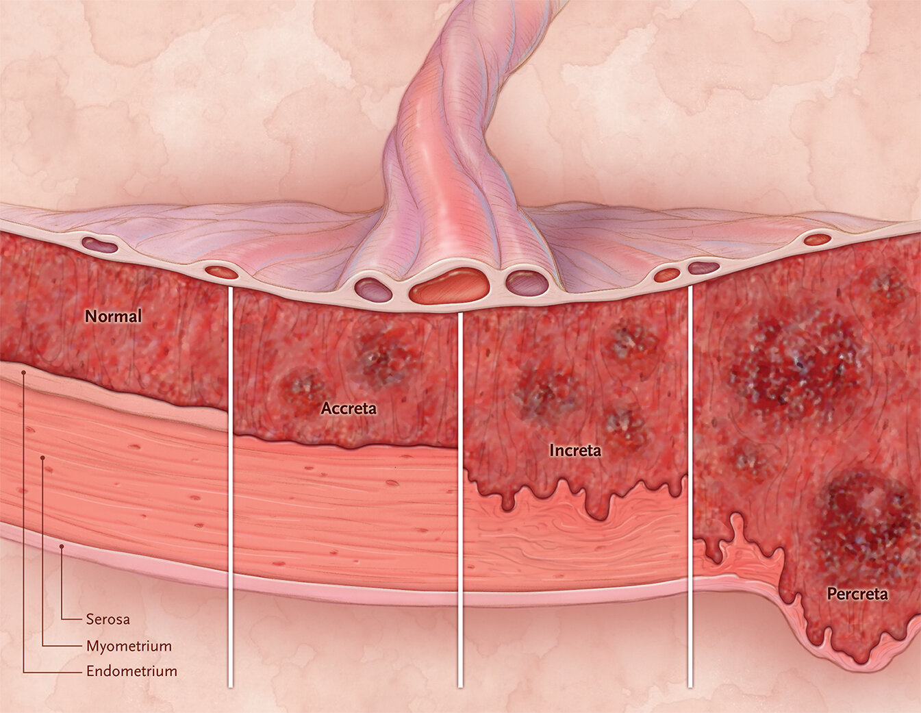
PAS has traditionally been thought of as an “invasive” disease, but that thinking is evolving to think of PAS as a disease of uterine dehiscence. The loss of the uterine decidua due to prior uterine scarring (i.e., due to surgery) brings about abnormal attachment and a “superhighway of vascularity,” thus that when delivery comes, the placenta fails to separate normally. Uterine muscle dehiscence likely accounts for the degree of invasiveness. It’s likely that cesarean scar pregnancies are a precursor to PAS.

https://resident360.nejm.org/clinical-pearls/placenta-accreta-spectrum

https://resident360.nejm.org/clinical-pearls/placenta-accreta-spectrum

The PAS diagnosis and terminology is also changing, from the traditional accreta / increta / percreta divide seen above, to a FIGO staging system with both surgical and pathologic criteria. You can review those here.

We review some of the risk factors, but far and away the biggest is a combination of a prior cesarean and placenta previa. Dr. Shainker mentions Dr. Robert Silver’s landmark paper on this - the percentages are worth committing to memory. Other risk factors include other types of uterine surgery like abdominal myomectomy; IVF and ART; and potentially dilation and curettage, though that is controversial.

Screen Shot 2021-03-06 at 08.11.09.png

Imaging is great in research capacities with high sensitivity (approaching 90+%), but only 50% of patients with accreta know about it before delivery. So the real world sensitivity is very poor. Risk factors should raise suspicion primarily, and the use of imaging help guide your preoperative suspicion. SMFM has now published a consensus on ultrasound diagnosis, which is the gold standard. More data should hopefully improve the real-world detection rates for PAS.

Cardiovascular Disease in Pregnancy Part III: Septal Defects and Maternal Congenital Cardiac Disease

We continue our series on cardiovascular diseases with part III this week. Check out Part I and Part II if you haven’t already!

Atrial Septal Defects (ASDs) + Ventricular Septal Defects (VSDs) 

  • ASD: Repaired vs. unrepaired 

    • Repaired = WHO class I - if no significant residual disease, have very low maternal cardiac risk in pregnancy.

    • Unrepaired = WHO class II 

      • Depends on size; if small → generally uncomplicated, will tolerate pregnancy well 

      • However, those with unrepaired ASDs also can have risk for supraventricular arrhythmias, like atrial flutter 

        • Remember that if there is an ASD, DVTs can travel to the systemic circulation → stroke (paradoxical embolus).

      • Also, if ASD is large and associated with significant pulmonary vascular disease, pregnancy should be avoided due to high risk of maternal and fetal mortality 

  • VSD: Repaired vs. unrepaired 

    • Repaired = WHO Class I. If small VSD with shunt ratio <1.7 with normal pulmonary pressure and preserved aerobic function → no increased risk to mom or fetus during pregnancy.

      • Shunt ratio briefly: pulmonary flow/systemic flow 

      • Basically, when there is L → R shunt, because there is a backleak of blood from the left side to the right side, the pulmonary flow will always be more than the systemic flow.

        • So a large shunt ratio implies significantly more pulmonary flow compared to systemic flow.

    •  Unrepaired: if large VSD shunts, history of arrhythmia associated with shunt, ventricular dysfunction, or pulmonary hypertension → higher risk of developing cardiovascular complications in pregnancy 

      • Complications include arrhythmias and heart failure.

      • Surveillance during pregnancy for pulmonary hypertension.

  • Atrioventricular septal defect 

    • After ASD repair, pregnancy is usually well tolerated (WHO Class II - III) 

    • Arrhythmias and worsening AV regurg have been described 

Tetralogy of Fallot 

  • Most common cyanotic congenital heart defect. Remember PROV:

    • Pulmonary stenosis (RV outflow tract obstruction)

    • RV Hypertrophy (concentric)

    • Overriding aorta 

    • VSD 

  • How to remember all this: 

    • Think this way: The aorta and the pulmonary artery trunk are next to each other in the heart. Aorta comes from the LV and the pulmonary artery from the RV. Imagine if the aorta just became really big and took over the real estate of the pulmonary artery. What has to happen? 

      • Overriding aorta 

      • Aorta takes over the real estate and therefore breaks through the ventricular septum → VSD 

      • The pulmonary artery is now super small, and therefore the pulmonic valve must be super small → RV outflow tract obstruction 

      • RV now has to work harder → RV hypertrophy 

        • So really, it’s just one thing that went wrong! 

  • Repaired Tet = WHO Class II - generally good outcomes if no severe hemodynamic abnormalities before pregnancy  

    • Complications can include arrhythmias (6.4%) and heart failure (2.4%) 

    • Fetal complications = premature delivery, SGA, recurrent CHD of any type, and very small risk of fetal and perinatal mortality (0.5% and 1.4% respectively) 

    • Remember that there is a higher risk of 22q11.2 microdeletion in offspring 

      • Approximately 15% of patients with ToF and other conotruncal defects have chromosome 22q11.2 microdeletion, and genetic testing should be offered in the prenatal setting 

    • Follow up every trimester by cards, but if severe pulmonary regurg, monthly or bimonthly follow up 

  • Unrepaired Tet = WHO Class III, and pregnancy is not recommended 

    • Would need close f/u with cardiology 

More rare stuff 

  • Ebstein’s Anomaly 

    • What is it: Tricuspid valve is placed too low on the right ventricle → enlargement of R atrium and non-functioning tricuspid valve. 

    • Again, if uncomplicated, pregnancy is well tolerated (Who Class II) 

    • But if there is cyanosis (usually due to ASD) or heart failure → counsel against pregnancy  

  • Transposition of the Great Arteries 

    • What it is: the left side of the heart pumps to the pulmonary artery and right side of the heart pumps to the aorta (basically, aorta and pulmonary artery are switched) 

    • Adults will have it corrected = arterial switch, and can usually tolerate pregnancy well if there is good clinical function pre-pregnancy 

    • Again, there is higher risk of heart failure and arrhythmias and should have good cardiology follow up 

  • Fontan Circulation 

    • What is it:

      • Basically it is a palliative surgical procedure performed in patients with a functional or anatomic single ventricle 

      • Some common reasons: hypoplastic left heart syndrome, tricuspid atresia, pulmonary atresia with intact ventricular septum, etc. 

    • Essentially diverts systemic venous return to the lungs without a pump, driven by central venous pressure. The single ventricle does pump blood to the systemic circulation.

    • For this to work, the person must have a low pulmonary arteriolar resistance, and relatively normal function of the single ventricle.

    • Prior to pregnancy, those with Fontan circulation should discuss with their cardiologist and have preconception counseling with MFM 

    • Those with poor functional capacity, history of heart failure, or ventricular function <40%, arrhythmias, etc should not get pregnant due to risk of complications 

    • Complications during pregnancy: arrhythmias, thrombotic and bleeding events, ventricular dysfunction, and edema 

    • Increased risk for SAB and premature birth as well as FGR 

    • PPH has been documented in up to 50% of pregnancies 

Labor and Delivery 

  • In most of these cases, patients should have telemetry intrapartum and 24 hours pp due to higher risk of arrythmias 

  • Strict I/Os

  • Depending on severity of case, but should consider early epidural

  • Vaginal delivery is not contraindicated, but should be assessed on a case by case basis

Intrahepatic Cholestasis of Pregnancy

Further reading for today: SMFM consult series #53 — it’s free!

And check out the ObG Project’s summary too!

What is cholestasis?

  • ICP or IHCP is a strange condition occurring in the 2nd and 3rd trimesters of pregnancy, and is characterized by two primary things: intense pruritis, and elevated serum bile acid levels.

    • The intense itching is the characteristic symptom -- rashless, unrelenting, and affecting even the palmar surfaces of the hands and the soles of the feet.

  • Women at risk of ICP include women who have had a history of ICP, women with hepatic disorders such as hepatitis C, nonalcoholic cirrhosis or pancreatitis, and women with gallstones and cholecystitis. 

    • Recurrence risk in subsequent pregnancy may be as high as 90%, but there’s limited data on this risk overall. 

    • ICP is also associated with multiple gestations, advanced maternal age, and there is likely a familial component to ICP as well. 

  • Incidence of ICP is around 0.3 - 0.5%, but has been reported as affecting up to 15% of pregnancies.

  • It seems silly to care about a disorder that causes itching in pregnancy, and ICP really doesn’t seem to affect the mother otherwise -- however, ICP is associated with severe adverse perinatal outcomes, particularly stillbirth, meconium-stained fluid, and preterm birth.

Many other conditions can cause itching in pregnancy!

  • Differential diagnoses include atopic eruption of pregnancy (AEP), polymorphic eruption of pregnancy (PEP), and pemphigoid gestationis (PG). 

    • AEP is associated with an eczematous rash that breaks out on the face, eyelids, neck, antecubal and popliteal fossa, trunk, and extremities -- think of the terrible eczema you saw in kids on your pediatrics rotation, but now put it on a pregnant woman!  

    • PEP, or it’s former name PUPPS (pruritic urticarial papules and plaques of pregnancy, or PUPPS), is also associated with a rash that often breaks out in plaques on the abdomen and thighs, but will spare the arms generally. This is the most common dermatosis of pregnancy.

    • Finally, PG is super rare, but as you might remember from your dermatology rotations or Step 1 studying, “pemphigus” is derived from  the Greek word meaning pustules. The characteristic skin change of this disorder are vesicles and bullae, and though rare, requires consultation with dermatology immediately!

    • There are even other conditions that can cause itching without a rash in pregnancy - renal failure, HIV, multiple sclerosis, and psychiatric disease include just a few of the broad differential. 

      • However, any patient with generalized pruritis in the 2nd or 3rd trimester should receive an evaluation for possible ICP.

How do we evaluate for ICP?

  • There shouldn’t be a rash!

    • The exam should also look for potential other hepatic causes that might raise bile acids - dark urine, jaundice or scleral icterus, abdominal pain and colic are not common with ICP.

  • Bile acids are the laboratory of choice to evaluate for cholestasis of pregnancy.

    • Total serum bile acids > 10 micromol/L is the general cutoff for diagnosis of ICP.

    • Some labs will ask for a fasting draw, which can be impractical for a pregnancy evaluation. Trials reviewing this have demonstrated that differences in random and fasting bile acid levels are pretty small in pregnant women, so feel free to draw away at the time of suspicion!

      • This also helps because many times, bile acid measurements occur at specialty labs and take a few days to return. 

  • Evaluation should also look at transaminases as these may also be elevated in ICP. 

What is the antenatal management of ICP?

  • The primary adverse outcome we’re trying to avoid is stillbirth. Stillbirth risk seems to correlate with increasing bile acid levels:

    • Diagnosis is called with BA > 10 micromol/L;

    • BA > 40 micromol/L has been demonstrated to have a higher incidence of stillbirth in some studies, as well as higher risk of other poor neonatal outcomes such as meconium, respiratory distress, and preterm birth; 

    • A large meta-analysis demonstrated the highest risk for stillbirth and other adverse outcomes seems to exist for women with BAs > 100 micromol/L. 

  • For maternal benefit, the drug of choice is ursodeoxycholic acid, or ursodiol

    • Ursodiol is effective in improving laboratory abnormalities (i.e., BAs, LFTs) and itching while not increasing any adverse fetal effects.

      • However, ursodiol likely does not decrease adverse fetal outcomes in ICP, so even though the bile acids will go lower, the peak value is still important to keep in mind. 

    • Dosing of ursodiol should be 10-15mg/kg per day, divided into 2-3 daily doses. 

      • This often comes out around 300mg BID or TID, or 500mg BID. 

    • Alternative medications for maternal benefit to reduce bile acid levels include cholestyramine and S-adenosyl-methionine (a dietary supplement in the US, sold as “SAMe”). 

    • Symptomatic treatment of itching can safely be targeted with oral antihistamines.

      • Topical antipruritics are likely unhelpful given the widespread nature of the itching!

    • Serial testing of bile acids is not necessarily recommended, but might be considered on a case-by-case basis as peak values are what have been reported in studies as being most significant with respect to stillbirth risk. 

      • It’s worthwhile to note that it’s not uncommon for pruritis in ICP can precede rising bile acid levels by even a few weeks! So in women who initially have a negative test, repeat bile acids and transaminase levels are very reasonable. 

        • Some may also practice in this particular scenario by diagnosing ICP clinically, and starting ursodiol empirically even in the absence of elevated bile acids. SMFM notes this is a reasonable approach, but certainly be aware of your differential diagnosis and keep an open eye for other pathologies.

  • For fetal benefit, our only defense against stillbirth is the use of antenatal testing! 

    • Antenatal testing should begin with diagnosis of ICP, at a gestational age when delivery would be performed in response to an abnormal test.

    • SMFM notes that most obstetric providers will perform some sort of antenatal testing, though the optimal frequency is unknown and even the effectiveness of testing is uncertain:

      • The mechanism for stillbirth in ICP is thought to be a sudden event, rather than a chronic placental vascular process -- so surveillance doesn’t necessarily predict stillbirth well.

    • Considerations for an antenatal testing schedule should be gestational age, severity of cholestasis (i.e., peak bile acid level), and patient values.

  • Ultimately to avoid stillbirth, delivery should be undertaken at an earlier gestational age:

    • In women with a clinical diagnosis of ICP (i.e., no bile acid elevation), delivery should not occur at less than 37 weeks.

    • In women with ICP and bile acids less than 100, delivery is reasonable between 36’0 - 39’0.

      • Data here is more challenging and less convincing on risk of stillbirth and when it occurs. It’s reasonable to consider managing women with lower values (ie., < 40) towards the later end of this window, and women with higher values (i.e., > 40) towards the earlier end.

    • In women with ICP and bile acids > 100, delivery can be offered as early as 36’0.

      • This is based on data suggesting in this high risk group, the risk of stillbirth substantially increases past this gestational age. 

      • SMFM even mentions in this group delivery between 34’0 - 36’0, provided that one of the following criteria is met:

        • Excruciating pruritis unremitting to pharmacotherapy;

        • History of ICP-associated stillbirth < 36 weeks in prior pregnancy;

        • Preexisting or acute hepatic disease with clinical or lab evidence of worsening hepatic function.

      • SMFM does advocate for the use of steroids for fetal lung maturity prior to delivery at 37’0 or earlier!